
Page 124 - Academic Press Encyclopedia of Physical Science and Technology 3rd BioTechnology
P. 124
P1: GLQ/GUB P2: FYK Final Pages
Encyclopedia of Physical Science and Technology EN007K-319 July 2, 2001 17:53
434 Hybridomas, Genetic Engineering of
cell fusion should be expected to secrete antibody. The
next stage involves selection of Mab-secreting hybrid-
omas from the population which has survived HAT treat-
ment. Cell clones can be isolated by the method of limiting
dilution. Cloning ensures that all cells in the cultures are
genetically identical. This involves dispensing a cell sus-
pension into a 96-well plate, so that each well contains an
average of one cell. Hybridomas grow poorly at low den-
sities but growth can be supported by feeder layer of cells.
This consists of a population of cells (e.g., thymocytes,
macrophages or splenocytes) which has been gamma ir-
radiated to prevent any growth. However, the metabolism
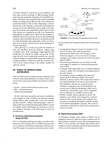
FIGURE 9 Enzyme-linked immunosorbent assay: ELISA.
continues and secreted growth factors can help the growth
of viable hybridoma cells particularly if they are inocu-
tions in which each component binds to the one previously
lated at low density. Feeder layers can be purchased as
added:
frozen suspensions.
After allowing 1–2 weeks for growth, the medium of
An appropriate antigen is bound to the plastic of the
each well should be tested for antibody content using
a suitable assay. Wells containing a high antibody titer base of the plate. Most large proteins bind
should then be selected for further cell growth. At this spontaneously. Difficulties with binding of small
stage the cells may be genetically unstable and a second molecular antigens can be overcome by forming a
round of cloning is recommended to ensure the isolation of conjugate with a larger molecule such as bovine serum
a stable population of high-level antibody-secreting cells. albumin (BSA).
This involves further testing of the culture medium for The remaining attachment sites are blocked on the
antibody content. solid support by addition of a noninterfering protein
such as BSA.
A solution of the Mab under test or a standard antibody
XII. ASSAY OF MONOCLONAL solution is added. This will bind to the antigen held on
ANTIBODIES
the solid support.
An antibody–enzyme conjugate with specificity
There are three assay procedures that are commonly used
against the first antibody is added. The second
to detect monoclonal antibodies in solution. Each is suit-
antibody is species specific. This means that it binds to
able for the measurement of Mab concentration in culture
any immunoglobulin of the species from which the
media.
first antibody is derived, e.g., goat anti-mouse
antibody. Conjugated to the second antibody is an
ELISA—enzyme-linked immunosorbent assay
enzyme such as alkaline phosphatase which can be
RIA—radioimmunoassay
detected by a colorimetric assay. These conjugated
Affinity chromatography
anti-Ig antibodies are available commercially.
Finally a suitable enzyme substrate is added. The
ELISA is the most commonly used assay for antibodies
substrate is one which can be changed to a colored
and is adapted to multi-well plates for analyzing multiple
product by the enzyme bound to the conjugate. For
samples. RIA is more sensitive but is more time consum-
example, p-nitrophenyl phosphate would be suitable
ing and expensive. Affinity chromatography is ideal if a
for an alkaline phosphatase conjugate. The extent of
high-performance liquid chromatography (HPLC) system
coloration in each well of a plate can be measured by a
is available and the hybridomas are growing in a serum-
multi-well reader.
free medium. The basis of the three types of assay are
described here:
B. Radioimmunoassay (RIA)
A. Enzyme-Linked Immunosorbent
A solid-phase binding assay similar to ELISA can be
Assay (ELISA)
adapted to radioactive detection when a radioactively la-
This is a solid-phase binding assay that can easily be per- beled antigen or antibody is used. The radioimmunoassay
formed in a 96-well plate (Fig. 9). ELISA measures anti- (RIA) is more sensitive and reliable than ELISA but is
gen or antibody concentration, depending on the protocol usually more time consuming and more expensive because
used. The stages of a typical assay involve a series of addi- of the cost of the radioactive label.