
Page 284 - Academic Press Encyclopedia of Physical Science and Technology 3rd BioTechnology
P. 284
P1: GPB/GRB P2: GLQ Final pages
Encyclopedia of Physical Science and Technology EN016J-783 August 1, 2001 10:58
Tissue Engineering 835
(usually less than 500 µm in diameter) with surfaces
treated to support cell attachment. These beads are then
maintained in suspension in medium using very low stir-
ring speeds in order to avoid mechanical cell damage,
either due to shearing forces in the liquid or due to bead–
bead collisions. The surface area available per microcar-
rier can be increased by using porous microcarriers, where
cells can migrate and proliferate within the porous matrix
as well as on the microcarrier surface; furthermore, cells
within the microcarrier are protected from mechanical
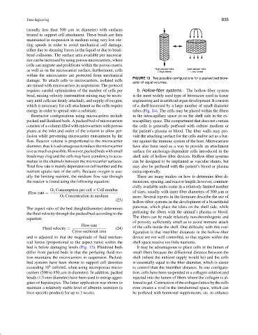
FIGURE 13 Two possible configurations for a packed bed biore-
damage. To attach cells to microcarriers, isolated cells
actor of equal volumes.
are mixed with microcarriers in suspension. The protocol
requires careful optimization of the number of cells per b. Hollow-fiber systems. The hollow-fiber system
bead, mixing velocity (intermittent mixing may be neces- is the most widely used type of bioreactor used in tissue
sary until cells are firmly attached), and supply of oxygen, engineering and in artificial organ development. It consists
which is necessary for cell attachment as the cells require of a shell traversed by a large number of small-diameter
energy in order to spread onto a substrate. tubes (Fig. 14). The cells may be placed within the fibers
Bioreactor configurations using microcarriers include in the intracapillary space or on the shell side in the ex-
packed and fluidized beds. A packed bed of microcarriers tracapillary space. The compartment that does not contain
consists of a column filled with microcarriers with porous the cells is generally perfused with culture medium or
plates at the inlet and outlet of the column to allow per- the patient’s plasma or blood. The fiber walls may pro-
fusion while preventing microcarrier entrainment by the vide the attaching surface for the cells and/or act as a bar-
flow. Reactor volume is proportional to the microcarrier rier against the immune system of the host. Microcarriers
diameter,thusitisadvantageoustoreducethemicrocarrier have also been used as a way to provide an attachment
size as much as possible. However, packed beds with small surface for anchorage-dependent cells introduced in the
beads may clog and the cells may have a tendency to accu- shell side of hollow-fiber devices. Hollow-fiber systems
mulate in the channels between the microcarrier surfaces. can be designed to be implanted as vascular shunts, but
Total flow rate is mainly dependent on cell number and the may also be perfused with the patient’s blood or plasma
nutrient uptake rate of the cells. Because oxygen is usu- extracorporeally.
ally the limiting nutrient, the medium flow rate through There are many studies on how to determine fiber di-
the reactor is found using the following equation: mensions, spacing, and reactor length; however, commer-
cially available units come in a relatively limited number
O 2 Consumption per cell × Cell number
Flow rate = of sizes, usually with inner fiber diameters of 500 µmor
O 2 Concentration in medium more. Several reports in the literature describe the use of
(23)
hollow-fiber systems in the development of a bioartificial
pancreas, which place the islets on the shell side, while
The aspect ratio of the bed (height/diameter) determines
perfusing the fibers with the animal’s plasma or blood.
the fluid velocity through the packed bed according to the
The fibers can be made relatively non-thrombogenic and
equation:
of porosity sufficiently small as to avoid immune attack
Flow rate
Fluid velocity = (24) of the cells inside the shell. One difficulty with this con-
Cross-sectional area figuration is that interfiber distances in the hollow-fiber
and is adjusted so that the magnitude of fluid mechan- device are not well controlled, so that regions within the
ical forces (proportional to the aspect ratio) within the shell space receive too little nutrients.
bed is below damaging levels (Fig. 13). Fluidized beds It may be advantageous to place cells in the lumen of
differ from packed beds in that the perfusing fluid mo- small fibers because the diffusional distance between the
tion maintains the microcarriers in suspension. Packed- shell (where the nutrient supply would be) and the cells
bed systems have been shown to support cell densities is essentially equal to the fiber diameter, which is easier
8
exceeding 10 cells/mL when using microporous micro- to control than the interfiber distance. In one configura-
carriers (500 to 850 µm in diameter). In addition, packed tion, cells have been suspended in a collagen solution and
beads (1.5-mm diameter) have been used to entrap aggre- injected into the lumen of fibers where the collagen is al-
gates of hepatocytes. The latter application was shown to lowedtogel.Contractionofthecollagenlatticebythecells
maintain a relatively stable level of albumin secretion (a even creates a void in the intraluminal space, which can
liver-specific product) for up to 3 weeks. be perfused with hormonal supplements, etc. to enhance