
Page 288 - Academic Press Encyclopedia of Physical Science and Technology 3rd BioTechnology
P. 288
P1: GPB/GRB P2: GLQ Final pages
Encyclopedia of Physical Science and Technology EN016J-783 August 1, 2001 10:58
Tissue Engineering 839
deposition of type II collagen and highly charged proteo- B. Epithelia and Endothelia
glycans, which are primarily responsible for the mechan-
1. Secretory and Transport Functions of
ical properties of native cartilage, is enhanced by subject-
Epithelial and Endothelial Cells
ing the tissue to cyclical mechanical compression. More
recently, chondrocytes seeded in polylactic–glycolic scaf- Epithelial and endothelial cells separate different com-
foldsandthenimplantedinectopicsitesinvivowerefound partments in the body; for example, endothelial cells sep-
to generate hyaline cartilage tissue with an overall shape arate the intravascular from tissue space, and intestinal
similar to that of the original synthetic matrix. Thus, it may epithelium separates the gut lumen from the inside of the
be possible to first generate the tissue of desired properties body. They control transport across these compartments,
at ectopic sites and, when ready, implant it at the site forming a selective barrier that prevents the transloca-
requiring intervention. tion of certain metabolites while favoring the transport
of others, sometimes through energy-dependent processes
(especially when the direction of transport is against the
2. In Vivo Regeneration Using
concentration gradient). In some cases, epithelial and en-
Guidance Templates
dothelial cells also perform important secretory functions,
Itissometimesmoreconvenienttopromotetissueregener- such as the release of antithrombogenic factors by en-
ation in situ, in which case the task of the tissue engineer dothelial cells and an array of secretory and biochemi-
is to favor wound healing and help the body overcome cal functions by liver hepatocytes. Although hepatocytes
some of its own limitations with respect to tissue regener- in vivo also perform transport functions and form a sep-
ation. The first regeneration templates that became widely arate bile canalicular network, all current approaches to
available are biodegradable meshes for the treatment of bioartificialliverdevelopmentessentiallyignorethisprop-
burn wounds. These templates are made of cross-linked erty due to the complexity of reproducing the in vivo ar-
collagen–glycosaminoglycan complexes and are applied rangement of hepatocytes in liver. Furthermore, it has been
to the wound site to favor regeneration of the skin dermis. hypothesized that the most important hepatic functions
The regenerated surface then provides a suitable substrate required for survival involve secretory and biochemical
for the attachment of skin epidermal cells (keratinocytes), functions that do not require a functional bile canalicular
which can be from an autologous skin graft obtained from network. A partial list of tissue engineered endothelial and
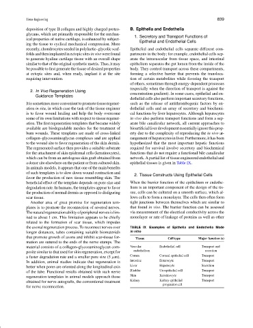
a donor site elsewhere on the patient or from cultured skin. epithelial tissues is given in Table IX.
In animals models, it appears that one of the main benefits
of such templates is to slow down wound contraction and 2. Tissue Constructs Using Epithelial Cells
favor the production of new tissue resembling skin. The
beneficial effect of the template depends on pore size and When the barrier function of the epithelium or endothe-
degradation rate. In humans, the templates appear to favor lium is an important component of the design of the tis-
the production of normal dermis as opposed to disfiguring sue, cells can be cultured on a smooth surface, which al-
scar tissue. lows cells to form a monolayer. The cells then often form
Another area of great promise for regeneration tem- tight junctions between themselves which are similar to
plates is to promote the reconnection of severed nerves. that found in vivo. The barrier function can be assessed
Thenaturalregenerationabilityofperipheralnervesislim- via measurement of the electrical conductivity across the
ited to about 1 cm. This limitation appears to be chiefly monolayer or rate of leakage of proteins as well as other
related to the formation of scar tissue, which impedes
the axonal regeneration process. To reconnect nerves over TABLE IX Examples of Epithelia and Endothelia Made
longer distances, tubes containing suitable biomaterials in vitro
that promote growth of axons and inhibit scar-tissue for- Tissue Cell type Major function (s)
mation are sutured to the ends of the nerve stumps. The
material consists of a collagen–glycosaminoglycan com- Vascular Endothelial cell Transport and
posite similar to that used for skin regeneration, except for endothelium secretion
a faster degradation rate and a smaller pore size (5 µm). Cornea Corneal epithelial cell Transport
In addition, animal studies indicate that regeneration is Intestine Enterocyte Transport
better when pores are oriented along the longitudinal axis Liver Hepatocyte Secretion
of the tube. Functional results obtained with such nerve Bladder Uroepithelial cell Transport
regeneration templates in animal models approach those Skin Keratinocyte Transport
obtained for nerve autografts, the conventional treatment Kidney Kidney epithelial Transport
progenitor cell
for nerve reconnection.