
Page 353 - Academic Press Encyclopedia of Physical Science and Technology 3rd Analytical Chemistry
P. 353
P1: GNH/FEE P2: GPJ Final Pages
Encyclopedia of Physical Science and Technology EN010C-493 July 19, 2001 20:30
718 Nuclear Magnetic Resonance (NMR)
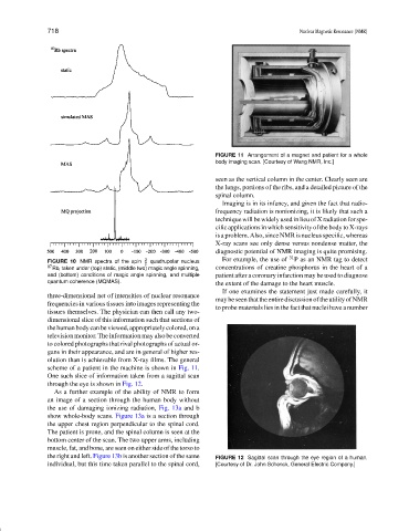
FIGURE 11 Arrangement of a magnet and patient for a whole
body imaging scan. [Courtesy of Wang NMR, Inc.]
seen as the vertical column in the center. Clearly seen are
the lungs, portions of the ribs, and a detailed picture of the
spinal column.
Imaging is in its infancy, and given the fact that radio-
frequency radiation is nonionizing, it is likely that such a
techniquewillbewidelyusedinlieuofXradiationforspe-
cific applications in which sensitivity of the body to X-rays
isaproblem.Also,sinceNMRisnucleusspecific,whereas
X-ray scans see only dense versus nondense matter, the
diagnostic potential of NMR imaging is quite promising.
FIGURE 10 NMR spectra of the spin 3 2 quadrupolar nucleus For example, the use of 31 P as an NMR tag to detect
87 Rb, taken under (top) static, (middle two) magic angle spinning, concentrations of creatine phosphorus in the heart of a
and (bottom) conditions of magic angle spinning, and multiple patient after a coronary infarction may be used to diagnose
quantum coherence (MQMAS).
the extent of the damage to the heart muscle.
If one examines the statement just made carefully, it
three-dimensional net of intensities of nuclear resonance
maybeseenthattheentirediscussionoftheutilityofNMR
frequencies in various tissues into images representing the
to probe materials lies in the fact that nuclei have a number
tissues themselves. The physician can then call any two-
dimensional slice of this information such that sections of
the human body can be viewed, appropriately colored, on a
television monitor. The information may also be converted
to colored photographs that rival photographs of actual or-
gans in their appearance, and are in general of higher res-
olution than is achievable from X-ray films. The general
scheme of a patient in the machine is shown in Fig. 11.
One such slice of information taken from a sagittal scan
through the eye is shown in Fig. 12.
As a further example of the ability of NMR to form
an image of a section through the human body without
the use of damaging ionizing radiation, Fig. 13a and b
show whole-body scans. Figure 13a is a section through
the upper chest region perpendicular to the spinal cord.
The patient is prone, and the spinal column is seen at the
bottom center of the scan. The two upper arms, including
muscle, fat, and bone, are seen on either side of the torso to
the right and left. Figure 13b is another section of the same FIGURE 12 Sagittal scan through the eye region of a human.
individual, but this time taken parallel to the spinal cord, [Courtesy of Dr. John Schenck, General Electric Company.]