
Page 354 - Academic Press Encyclopedia of Physical Science and Technology 3rd Analytical Chemistry
P. 354
P1: GNH/FEE P2: GPJ Final Pages
Encyclopedia of Physical Science and Technology EN010C-493 July 19, 2001 20:30
Nuclear Magnetic Resonance (NMR) 719
FIGURE 13 (a) Scan through upper trunk perpendicular to spine. (b) Scan through upper trunk parallel to the spinal
cord. [Courtesy of Dr. John Schenck, General Electric Company.]
of characteristic fingerprints that can be used to probe their ation times available to the nucleus. The field of imaging is
environments. We have talked about fingerprints associ- just beginning to use these fingerprints for enhanced reso-
ated with shielding, dipolar interactions, quadrupolar in- lution. For example, there now exist “T 1 images”, and “T 2
teractions, and their reflections as seen in the various relax- images” that use the fact that transverse and longitudinal
relaxation times of nuclei in a given tissue are characteris-
tic of that tissue. The full range of interactions of nuclear
behavior has yet to be exploited for imaging.
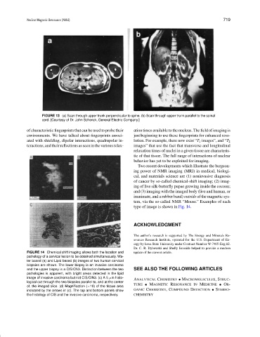
Two recent developments which illustrate the burgeon-
ing power of NMR imaging (MRI) in medical, biologi-
cal, and materials science are (1) noninvasive diagnosis
of cancer by so-called chemical-shift imaging; (2) imag-
ing of live silk-butterfly pupae growing inside the cocoon;
and (3) imaging with the imaged body (live and human, or
inanimate, and a rubber band) outside of the magnetic sys-
tem, via the so-called NMR “Mouse.” Examples of each
type of image is shown in Fig. 14.
ACKNOWLEDGMENT
The author’s research is supported by The Energy and Minerals Re-
sources Research Institute, operated for the U.S. Department of En-
ergy by Iowa State University under Contract Number W-7405-Eng.82.
Dr. C. R. Dybowski and Shelly Ironside helped to provide a modern
FIGURE 14 Chemical shift imaging allows both the location and update of the current article.
pathology of a cervical lesion to be obtained simultaneously. Wa-
ter based (a) and Lipid based (b) images of two human cervical
biopsies are shown. The lower biopsy is an invasive carcinoma
and the upper biopsy is a CIS/CN3. Distinction between the two SEE ALSO THE FOLLOWING ARTICLES
pathologies is apparent, with bright areas detected in the lipid
image of invasive carcinoma but not CIS/CIN3. (c) A 5 µm histo- ANALYTICAL CHEMISTRY • MACROMOLECULES,STRUC-
logical cut through the two biopsies parallel to, and at the center
of, the imaged slice. (d) Magnification (×10) of the tissue area TURE • MAGNETIC RESONANCE IN MEDICINE • OR-
indicated by the arrows in (c). The top and bottom panels show GANIC CHEMISTRY,COMPOUND DETECTION • STEREO-
the histology of CIS and the invasive carcinoma, respectively. CHEMISTRY