
Page 69 - Academic Press Encyclopedia of Physical Science and Technology 3rd BioChemistry
P. 69
P1: GTQ/GUU P2: GLM Final Pages
Encyclopedia of Physical Science and Technology EN008K-353 June 29, 2001 12:41
102 Ion Transport Across Biological Membranes
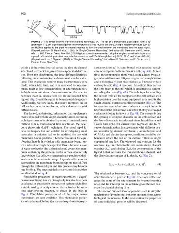
FIGURE 3 The single-channel current-recording technique. (A) The tip of a borosilicate glass pipet, with a tip
opening of 1–2 µm is pressed against the membrane of a frog muscle cell (left). A slight negative pressure (20–30
cm H 2 O) is applied to the pipet for several seconds to form the seal between the membrane and the pipet (right).
(Reproduced from O. Hamill et al. (1995). In “Single-Channel Recording,” 2nd edition (B. Sakmann and E. Neher,
eds.), p. 663, Plenum Press, New York.) (B) A typical current trace recorded using the single-channel technique, a rat
◦
myoball cell containing nicotinic acetylcholine receptors, and 20-µM acetylcholine (pH 7.2, 22 C, and V m =−80 mV).
(Reproduced from F. Sigworth (1983). In “Single-Channel Recording,”first edition (B. Sakmann and E. Neher, eds.),
Plenum Press, New York.)
within a definite time interval versus the time the channel carbamoylcholine] is equilibrated with nicotine acetyl-
was closed is expected to give a three-exponential distribu- choline receptors on the surface of a cell (Fig. 4A). At zero
tion. From this distribution, the three different lifetimes, time, the compound is photolyzed, using a laser, by a sin-
reflecting the constants to be determined, can be calcu- gle pulse within about 100 µsec to give carbamoylcholine
lated. This evaluation requires many measurements to be and a biologically inert side-product, a 2-nitroso-α-keto
made, which take time, and it is restricted to measure- carboxylic acid (Fig. 4 reaction). An optical fiber carries
ments made at low concentrations of neurotransmitters. the light beam to the cell, which is attached to a current-
At higher concentrations of neurotransmitter, the receptor recording electrode (Fig. 4A). The technique for recording
becomes inactive, desensitized (in the millisecond time the current from all the receptors on the cell surface with
region) (Fig. 2) and the signal to be measured disappears. high precision uses the same equipment as is used in the
Additionally, we now know that many receptors on the single-channel current-recording technique (Fig. 3). The
cell surface exist in two forms, which desensitize with increase in current that results when carbamoylcholine is
different rates. liberated on the cell surface, due to the photolysis of caged
The desired, and missing, information that supplements carbamoylcholine is shown in Fig. 4B. The current is due
results obtained with thesingle-channel current–recording the opening of receptor-channels on the cell surface and
technique can now be obtained by using a transient kinetic the flow of inorganic ions through them. In a different and
method with a microsecond time resolution, the laser- slower time zone, the current then decreases due to re-
pulse photolysis (LaPP) technique. The usual rapid ki- ceptor desensitization. In experiments with different neu-
netic techniques that are suitable for investigating small rotransmitter [glutamate, serotonin, γ -aminobutyric acid
molecules in solution had to be modified for use with (GABA), and glycine] receptors, conditions could be ob-
membrane-bound proteins. The time resolution for equi- tained in which the rise of the current follows a single
librating ligands in solution with membrane-bound pro- exponential rate law. The observed rate constant for the
teins is less than might be expected. This is because a layer rise time, k obs , is related to the rate constants for channel
of water molecules (the diffusion layer) covers the mem- opening (k op ) and closing (k cl ), the concentration of the
brane containing the proteins on the surface of relatively ligand L that activates the transmembrane channel, and
large objects like cells, or even membrane patches with di- the dissociation constant of L, that is, K 1 (Fig. 2):
ameters in the micrometer range. Ligands in the solution
surrounding the membrane-bound receptors must diffuse 2
k obs = k cl + k op [L/(L + K 1 )] . (4)
through the diffusion layer and this process may become
rate limiting. The steps needed to overcome this problem
are illustrated in Fig. 4. The relationship between k obs and the concentration of
Photolabile precursors of neurotransmitters (“caged” neurotransmitter is given in Fig. 4C. The slope of the line
neurotransmitters) that are biologically inactive have been gives the value of the rate constant for channel opening
developed. A photolabile precursor of carbamoylcholine, (k op ) and the intercept on the ordinate gives the rate con-
a stable analog of acetylcholine that activates the nico- stant for channel closing (k cl ).
tinic acetylcholine receptor, is shown in the inset to This section outlined some approaches used to study the
Fig. 4. Photolabile precursors of all the major neuro- mechanism of proteins that transport inorganic ions across
transmitters are now available. This photolabile precur- biological membranes. In the next section the properties
sor of carbamoylcholine ([N-(α-carboxy-2-nitrobenzyl)- of some individual proteins will be discussed.