
Page 71 - Academic Press Encyclopedia of Physical Science and Technology 3rd BioChemistry
P. 71
P1: GTQ/GUU P2: GLM Final Pages
Encyclopedia of Physical Science and Technology EN008K-353 June 29, 2001 12:41
104 Ion Transport Across Biological Membranes
V m at about −40 mV. (7) Lowering the concentration of
cGMP in the cell lowers the concentration of open Na -
+
conducting channels, which to remain open must have
cGMP bound to them. (8) The closing of cGMP-activated
transmembrane channels decreases the flux rate of Na +
into the cell and, therefore, changes the voltage across the
membrane. (9) The changes in V m result in the release
by the retinal cell of chemical signals (neurotransmitters)
adjacent to another cell. (10) The neurotransmitters bind
to receptors on an adjacent cell that transiently form trans-
membrane channels, allowing cations or anions, depend-
ing on the receptor, to move through the cell membrane.
IV. INORGANIC ION TRANSPORT AND
INTEGRATION OF ENVIRONMENTAL
INFORMATION
In the previous section, the transport of ions across the
membrane was initiated by an environmental signal,
namely, light. This resulted in a change in the transmem-
brane voltage and a subsequent influx of calcium ions into
the nerve terminal of a sensory cell (Fig. 1), resulting in
the release of a chemical signal. In general, these chemical
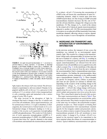
FIGURE 5 (A) Light-induced transformation of 11-cis-retinal to signals (neurotransmitters) are released from the nerve
all-trans-retinal in visual pigments. 11-Cis-retinal is attached terminals of one cell and diffuse across a gap between cells
through a Schiff-base linkage of lysine 256 in rhodopsin. (B) called a synapse (Fig. 1). In the membrane of the adjacent
Rhodopsin has a molecular weight of 40,000. Seven transmem-
cell, about 20–40 nm removed from the nerve terminal,
brane helices are embedded in the cell membranes. 11-Cis-retinal
lies near the center of the lipid membrane. The structure is based the neurotransmitters bind to transmembrane neurotrans-
on the three-dimensional reconstruction of electron microscope mitter receptors. On binding the neurotransmitter, these
images by Henderson and Unwin. (Reproduced with permission transmembrane proteins, transiently (a few milliseconds)
from Nelson, D. L., and Cox, M. M. p. 460. Figs. 13–22. “Lehninger open a transmembrane channel (Fig. 1). These channels
Principles of Biochemistry” (2000). 3rd edition, Worth Publishers are specific for inorganic cations, sodium, or potassium,
p. 460.)
and some are also specific for calcium or chloride ions,
depending on the receptor. If the resulting change in the
light, leads to the release of all-trans-retinal. The all-trans- transmembrane voltage, V m , is of appropriate sign and
retinal is transformed to all-trans-retinol (Vitamin A) by magnitude (∼+ 20 mV) the voltage change will be prop-
pigment epithelia cells in the eye. It is a precursor in the agated along the axon of the cell (Fig. 1), by a process in
synthesis of 11-cis-retinal, which is then transported back which sodium and potassium ions move across the mem-
to cells containing opsin and reattached to the ∈-amino brane. This is described in detail at the end of this section.
group of lysine 296 of opsin. The major neurotransmitters are listed in Table I. Typ-
The steps involved in the absorption of light by ical excitatory neurotransmitters are acetylcholine, gluta-
rhodopsin that lead to changes in the flux of sodium ions mate, and serotonin. They bind to receptors that are named
across the cell membrane, and to signal transmission, can in the same way, for instance, the acetylcholine receptor,
be summarized as follows. (1) Light is absorbed. (2) Sub- glutamate receptor, etc. These receptors on binding their
sequently, the isomerization of retinal results in (3) a con- specific neurotransmitter form transmembrane channels
formational change of rhodopsin; leading to (4) the ac- specific for the inorganic sodium and potassium cations.
tivation of the enzyme cyclic-guanosine monophosphate They are called excitatory receptors because they shift the
(cGMP) phosphodiesterase. This results in (5) the hydrol- transmembrane potential of the cell membrane to more
ysis of cyclic guanosine monophosphate (cGMP) to 5 positive values. A change in V m of about +20 mV is re-
guanosine monophosphate (5 -GMP). (6) cGMP activates quired for the electrical signal to be transmitted to the
a protein that forms Na -specific channels in the retinal nerve terminal where the release of a neurotransmitter is
+
cell membrane and maintains the transmembrane potential elicited. Typical inhibitory neurotransmitters are glycine