
Page 97 - Academic Press Encyclopedia of Physical Science and Technology 3rd BioChemistry
P. 97
P1: GST/MBQ P2: GQT Final Pages
Encyclopedia of Physical Science and Technology EN009G-417 July 10, 2001 15:10
358 Membrane Structure
Sphingomyelin hydrolysis yields ceramide, a lipid me- For PC, PE, and PG, the glycerol backbone is oriented
diator involved in regulating cell growth, cell differenti- perpendicular to the bilayer surface, while the polar head-
ation, and cell death. Glycosphingolipids act as specific groups are almost parallel to the membrane surface. Neu-
recognition sites in eukaryotic cells, and they determine tron scattering experiments of selectively deuterated lipid
blood-group, organ, and tissue specificity and are further headgroups in liquid crystalline and gel state membranes
involved in tissue immunity and cell–cell recognition. determine the mean label position with an accuracy of up
˚
The phospholipids found in prokaryotic (bacterial) and to ±1 A and provide independent support for the almost
eukaryotic (mammalian) cell membranes usually contain parallel headgroup orientation of PC, PE, and PG.
saturated as well as cis-unsaturated fatty acyl chains, the The headgroup orientations of PC, PE, and PG bilayers
most abundant being the saturated palmitoyl (C 16:0) and in the liquid crystalline phase, in the gel phase, and in sin-
the cis-unsaturated oleoyl chains (C 18:1, cis). There is gle crystals are thus very similar and independent of the
a strong positional preference for the two types of fatty dynamic state of the membrane. The correlation times of
acids, with the saturated and the unsaturated chain being the segmental and collective motions of the head groups
localized at positions 1 and 2, respectively, of the glyc- decrease abruptly by more than two orders of magnitude
erol backbone of the lipid molecule. Phospholipids with at the gel-to-liquid phase transition; nevertheless, the av-
a single cis-double bond are predominant, but lipids con- erage conformation remains unaltered.
taining more than one double bond also occur quite com- Phosphatidylserine measured at neutral pH and in the
monly. In membranes of the nervous system, polyunsat- absence of ions is similar to the other phospholipids with
urated fatty acids appear to be critical for proper mem- respect to the glycerol backbone, but differs distinctly in
brane functioning. The length of the fatty acyl chains its headgroup orientation and motion. The PS headgroup
and the degree of chain unsaturation as well as the size, is rigid and exhibits little internal flexibility. A crystal
the charge, and the hydrogen-bonding capacity of head- structure is not available so far.
groups determine the intermolecular lipid–lipid interac- For the comparison of NMR and X-ray diffraction mea-
tions reflected in the lipid packing density and the gel- surements, the effect of membrane hydration can be rele-
to-liquid crystal phase transition temperatures of lipid vant. A minimum of 11 to 16 water molecules per lipid
membranes. The effect of headgroups on the gel-to-liquid molecule is needed to form a primary hydration shell for
crystal phase transition temperature, T c , is illustrated PC, PE, and PG. Additional water is in exchange with the
by the following series of lipids (C 16:0) mixed with water: primary hydration shell. With increasing hydration (10–
+
−
◦
◦
◦
PC (T c = 41 C) ∼ PG (T c = 41 C) ∼ SM (T c = 41 C) < 70 wt% H 2 O) the −P -N dipole of the phosphocholine
◦
PS deprotonated (T c = 54 C) < PS protonated (T c = headgroup was shown to move with its cationic end away
62 C) < PE (T c = 64 C) < PA (T c = 71 C) < GalCer from the hydrocarbon layer. This explains why the −P -
◦
−
◦
◦
(T c = 82 C). Knowledge of the gel-to-liquid crystal phase N dipoles in liquid crystalline membranes are generally
+
◦
transitions is of relevance for the question of lipid domain slightly tilted away from the membrane surface up to an
formation to be discussed below. angle of about 30 , while they are oriented parallel to the
◦
surface in the crystal structure.
III. THE MEMBRANE–WATER INTERFACE
B. Ester Linkage of the sn-2 Fatty Acyl Chain
The membrane–water interface comprises the lipid head-
Is Part of the Lipid–Water Interface
group proper, with tightly bound hydration water and a
more loosely packed extended hydration layer; the glyc- If the two fatty acyl chains of lipids are deuterated at
erol backbone; and the ester linkages of the fatty acyl methylene groups immediately next to the ester link-
chains. Due to conformational constraints, the carbonyl age they give rise to quite different quadrupole split-
group of the sn-2 chain is particularly close to the lipid– tings, indicating that the beginnings of the two chains
water interface, while that of the sn-1 chain is inserted have different conformations. The inequivalence of the
deeper into the membrane interior. two chains was first observed for 1,2-dipalmitoyl-sn-
glycero-3-phosphocholine (DPPC) in its liquid crys-
talline phase. It is also preserved in the presence of
A. Headgroup and Glycerol Backbone
transmembrane proteins in reconstituted and in natural
Conformation of Phospholipids
membranes.
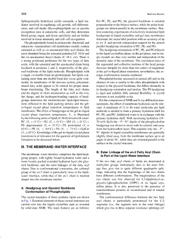
The crystal structures of three synthetic lipids are shown The conformational difference between the two fatty
in Fig. 1. Essential elements of these crystal structures are acyl chains is particularly pronounced for the C-2
carried over into the liquid crystalline state as revealed segments (i.e., the segment next to the ester linkage)
by solid-state NMR. The main features are as follows: which give rise to three separate resonance splittings (cf.