
Page 101 - Academic Press Encyclopedia of Physical Science and Technology 3rd BioChemistry
P. 101
P1: GST/MBQ P2: GQT Final Pages
Encyclopedia of Physical Science and Technology EN009G-417 July 10, 2001 15:10
362 Membrane Structure
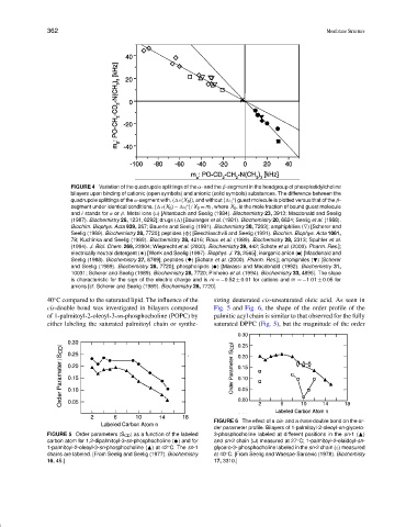
FIGURE 4 Variation of the quadrupole splittings of the α- and the β-segment in the headgroup of phosphatidylcholine
bilayers upon binding of cationic (open symbols) and anionic (solid symbols) substances. The difference between the
◦
quadrupole splittings of the α-segment with, ( ν(X b )), and without ( ν ) guest molecule is plotted versus that of the β-
i
segment under identical conditions, ( ν(X b ) − ν )/X b = m i , where X b , is the mole fraction of bound guest molecule
◦
i
and i stands for α or β. Metal ions ( ) [Altenbach and Seelig (1984). Biochemistry 23, 3913; Macdonald and Seelig
(1987). Biochemistry 26, 1231, 6292]; drugs ( ) [Boulanger et al. (1981). Biochemistry 20, 6824; Seelig et al. (1988).
Biochim. Biophys. Acta 939, 267; Bauerle and Seelig (1991). Biochemistry 30, 7203]; amphiphilies (∇) [Scherer and
Seelig (1989). Biochemistry 28, 7720]; peptides (
) [Beschiaschvili and Seelig (1991). Biochim. Biophys. Acta 1061,
?
78; Kuchinka and Seelig (1989). Biochemistry 28, 4216; Roux et al. (1989). Biochemistry 28, 2313; Spuhler et al.
(1994). J. Biol. Chem. 269, 23904; Wieprecht et al. (2000). Biochemistry 39, 442; Schote et al. (2000). Pharm. Res.];
electrically neutral detergent (* ) [Wenk and Seelig (1997). Biophys. J. 73, 2565]; inorganic anion ( ) [Macdonald and
Seelig (1988). Biochemistry 27, 6769]; peptides ( ) [Schote et al. (2000). Pharm. Res.]; amphiphiles ( ) [Scherer
and Seelig (1989). Biochemistry 28, 7720]; phospholipids ( ✉ ) [Marassi and Macdonald (1992). Biochemistry 31,
10031; Scherer and Seelig (1989). Biochemistry 28, 7720; Pinheiro et al. (1994). Biochemistry 33, 4896]. The slope
is characteristic for the sign of the electric charge and is m =−0.52 ± 0.01 for cations and m =−1.01 ± 0.05 for
anions [cf. Scherer and Seelig (1989). Biochemistry 28, 7720].
◦
40 C compared to the saturated lipid. The influence of the sizing deuterated cis-unsaturated oleic acid. As seen in
cis-double bond was investigated in bilayers composed Fig. 5 and Fig. 6, the shape of the order profile of the
of 1-palmitoyl-2-oleoyl-3-sn-phosphocholine (POPC) by palmitic acyl chain is similar to that observed for the fully
either labeling the saturated palmitoyl chain or synthe- saturated DPPC (Fig. 5), but the magnitude of the order
FIGURE 6 The effect of a cis- and a trans-double bond on the or-
der parameter profile. Bilayers of 1-palmitoyl-2-oleoyl-sn-glycero-
FIGURE 5 Order parameters |S CD | as a function of the labeled 3-phosphocholine labeled at different positions in the sn-1 ( )
carbon atom for 1,2-dipalmitoyl-3-sn-phosphocholine ( ✉ ) and for and sn-2 chain ( ) measured at 27 C; 1-palmitoyl-2-elaidoyl-sn-
◦
1-palmitoyl-2-oleoyl-3-sn-phosphocholine ( ) at 42 C. The sn-1 glycero-3- phosphocholine labeled in the sn-2 chain (♦) measured
◦
chains are labeled. [From Seelig and Seelig (1977). Biochemistry at 40 C. [From Seelig and Waespe-Sarcevic (1978). Biochemisty
◦
16, 45.] 17, 3310.]