
Page 41 - Academic Press Encyclopedia of Physical Science and Technology 3rd InOrganic Chemistry
P. 41
P1: ZBU Final Pages
Encyclopedia of Physical Science and Technology EN002F-55 May 22, 2001 21:6
Bioinorganic Chemistry 129
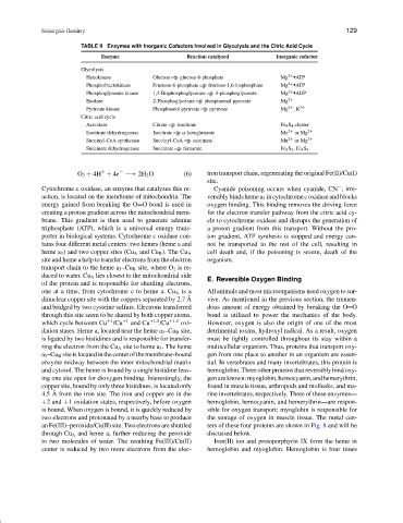
TABLE II Enzymes with Inorganic Cofactors Involved in Glycolysis and the Citric Acid Cycle
Enzyme Reaction catalyzed Inorganic cofactor
Glycolysis
Hexokinase Glucose glucose-6-phosphate Mg 2+ • ATP
Phosphofructokinase Fructose-6-phosphate fructose-1,6-bisphosphate Mg 2+ • ATP
Phosphoglycerate kinase 1,3-Bisphosphoglycerate 3-phosphoglycerate Mg 2+ • ADP
Enolase 2-Phosphoglycerate phosphoenol pyruvate Mg 2+
Pyruvate kinase Phosphoenol pyruvate pyruvate Mg 2+ ,K 2+
Citric acid cycle
Aconitase Citrate isocitrate Fe 4 S 4 cluster
Isocitrate dehydrogenase Isocitrate α-ketoglutarate Mn 2+ or Mg 2+
Succinyl-CoA synthetase Succinyl-CoA succinate Mn 2+ or Mg 2+
Succinate dehydrogenase Succinate fumarate Fe 2 S 2 ,Fe 4 S 4
−
+
O 2 + 4H + 4e −→ 2H 2 O. (6) tron transport chain, regenerating the original Fe(II)/Cu(I)
site.
Cytochrome c oxidase, an enzyme that catalyzes this re- Cyanide poisoning occurs when cyanide, CN , irre-
−
action, is located on the membrane of mitochondria. The versibly binds heme a 3 in cytochrome c oxidase and blocks
energy gained from breaking the O O bond is used in oxygen binding. This binding removes the driving force
creating a proton gradient across the mitochondrial mem- for the electron transfer pathway from the citric acid cy-
brane. This gradient is then used to generate adenine cle to cytochrome oxidase and disrupts the generation of
triphosphate (ATP), which is a universal energy trans- a proton gradient from this transport. Without the pro-
porter in biological systems. Cytochrome c oxidase con- ton gradient, ATP synthesis is stopped and energy can-
tains four different metal centers: two hemes (heme a and not be transported to the rest of the cell, resulting in
cell death and, if the poisoning is severe, death of the
heme a 3 ) and two copper sites (Cu A and Cu B ). The Cu A
site and heme a help to transfer electrons from the electron organism.
transport chain to the heme a 3 –Cu B site, where O 2 is re-
duced to water. Cu A lies closest to the mitochondrial side
E. Reversible Oxygen Binding
of the protein and is responsible for shuttling electrons,
one at a time, from cytochrome c to heme a. Cu A is a All animals and most microorganisms need oxygen to sur-
˚
dinuclear copper site with the coppers separated by 2.7 A vive. As mentioned in the previous section, the tremen-
and bridged by two cysteine sulfurs. Electrons transferred dous amount of energy obtained by breaking the O O
through this site seem to be shared by both copper atoms, bond is utilized to power the mechanics of the body.
+1
which cycle between Cu /Cu +1 and Cu +1.5 /Cu +1.5 oxi- However, oxygen is also the origin of one of the most
dation states. Heme a, located near the heme a 3 –Cu B site, detrimental toxins, hydroxyl radical. As a result, oxygen
is ligated by two histidines and is responsible for transfer- must be tightly controlled throughout its stay within a
ring the electron from the Cu A site to heme a 3 . The heme multicellular organism. Thus, proteins that transport oxy-
a 3 –Cu B siteislocatedinthecenterofthemembrane-bound gen from one place to another in an organism are essen-
enzyme midway between the inner mitochondrial matrix tial. In vertebrates and many invertebrates, this protein is
and cytosol. The heme is bound by a single histidine leav- hemoglobin. Three other proteins that reversibly bind oxy-
ing one site open for dioxygen binding. Interestingly, the genareknown:myoglobin,hemocyanin,andhemerythrin,
copper site, bound by only three histidines, is located only found in muscle tissue, arthropods and mollusks, and ma-
˚
4.5 A from the iron site. The iron and copper are in the rine invertebrates, respectively. Three of these enzymes—
+2 and +1 oxidation states, respectively, before oxygen hemoglobin, hemocyanin, and hemerythrin—are respon-
is bound. When oxygen is bound, it is quickly reduced by sible for oxygen transport; myoglobin is responsible for
two electrons and protonated by a nearby base to produce the storage of oxygen in muscle tissue. The metal cen-
an Fe(III)–peroxide/Cu(II) site. Two electrons are shuttled ters of these four proteins are shown in Fig. 8 and will be
through Cu A and heme a, further reducing the peroxide discussed below.
to two molecules of water. The resulting Fe(III)/Cu(II) Iron(II) ion and protoporphyrin IX form the heme in
center is reduced by two more electrons from the elec- hemoglobin and myoglobin. Hemoglobin is four times