
Page 48 - Academic Press Encyclopedia of Physical Science and Technology 3rd Molecular Biology
P. 48
P1: GPB Final Pages
Encyclopedia of Physical Science and Technology EN004L-956 June 9, 2001 21:7
DNA Testing in Forensic Science 599
translated from 5 to 3 it is at the very end of the primer devices that scan a gel after the DNA products have
and should not affect the amplification or binding of the been separated by a process called electrophoreses. Ex-
primer if made correctly. One of the oldest fluorescent amples of post electrophoresis scanners are the Hitachi
®
dyes is fluoroscein. Many devices have been made that FMBIO Fluorescent Scanner, the Molecular Dynamics
will detect fluoroscein, and it has been used extensively to Fluorimager TM and the Beckman Genomics SC scanner.
label antibodies and other materials. Many of these dyes The Hitachi and Molecular Dynamics use a laser as a light
have been used for a long time and are in the public do- with filters to identify the proper frequency and a CCD
main. Others have been developed for specific projects. camera to capture the image. The Beckman Genomics SC
The dyes used by PE Biosystems were originally propri- uses a monochromatic Xenon light source and uses filters
etary and part of a patented four color DNA sequencing to detect the appropriate light for the CCD camera. The
system (Blue, Green, Yellow, Red). These dyes are now CCD camera scans back and forth over the gel as it is ex-
becoming more readily available. posedtothelightsourceanddetectsthevariousfluorescent
colors using filters that change. This type of equipment
has flexibility because different formats of electrophore-
B. Fluorescent Detection Equipment
sis gels can be used and scanned. The output is in the form
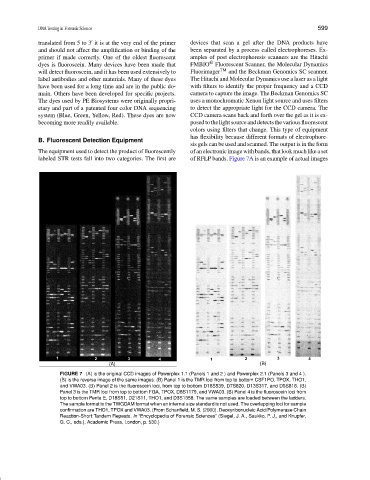
The equipment used to detect the product of fluorescently of an electronic image with bands, that look much like a set
labeled STR tests fall into two categories. The first are of RFLP bands. Figure 7A is an example of actual images
FIGURE 7 (A) is the original CCD images of Powerplex 1.1 (Panels 1 and 2 ) and Powerplex 2.1 (Panels 3 and 4 ).
(B) is the reverse image of the same images. (B) Panel 1 is the TMR loci from top to bottom CSF1PO, TPOX, THO1,
and VWA03. (B) Panel 2 is the fluoroscein loci, from top to bottom D16S539, D7S820, D13S317, and D5S818. (B)
Panel 3 is the TMR loci from top to bottom FGA, TPOX, D8S1179, and VWA03. (B) Panel 4 is the fluoroscein loci from
top to bottom Penta E, D18S51, D21S11, THO1, and D3S1358. The same samples are loaded between the ladders.
The sample format is the TWGDAM format when an internal size standard is not used. The overlapping loci for sample
confirmation are THO1, TPOX and VWA03. (From Schanfield, M. S. (2000). Deoxyribonucleic Acid/Polymerase Chain
Reaction-Short Tandem Repeats. In “Encyclopedia of Forensic Sciences” (Siegel, J. A., Saukko, P. J., and Knupfer,
G. C., eds.), Academic Press, London, p. 530.)