
Page 98 - Academic Press Encyclopedia of Physical Science and Technology 3rd Molecular Biology
P. 98
P1: GTQ Final pages
Encyclopedia of Physical Science and Technology EN017F-788 August 3, 2001 16:27
Translation of RNA to Protein 39
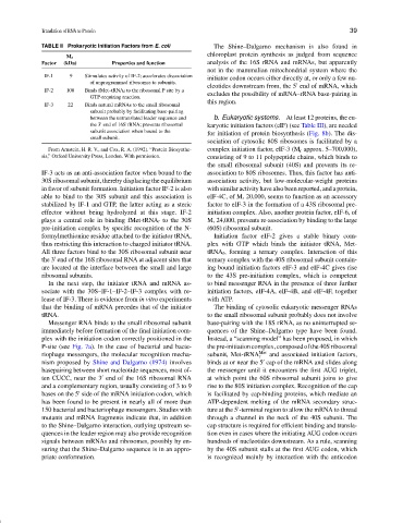
TABLE II Prokaryotic Initiation Factors from E. coli The Shine–Dalgarno mechanism is also found in
chloroplast protein synthesis as judged from sequence
M r
Factor (kDa) Properties and function analysis of the 16S rRNA and mRNAs, but apparently
not in the mammalian mitochondrial system where the
IF-1 9 Stimulates activity of IF-2; accelerates dissociation
initiator codon occurs either directly at, or only a few nu-
of unprogrammed ribosomes to subunits.
cleotides downstream from, the 5 end of mRNA, which
IF-2 100 Binds fMet–tRNA f to the ribosomal P site by a
excludes the possibility of mRNA–rRNA base-pairing in
GTP-requiring reaction.
this region.
IF-3 22 Binds natural mRNAs to the small ribosomal
subunit probably by facilitating base-pairing
between the untranslated leader sequence and b. Eukaryotic systems. At least 12 proteins, the eu-
the 3 end of 16S rRNA; prevents ribosomal karyotic initiation factors (eIF) (see Table III), are needed
subunit association when bound to the for initiation of protein biosynthesis (Fig. 8b). The dis-
small subunit.
sociation of cytosolic 80S ribosomes is facilitated by a
From Arnstein, H. R. V., and Cox, R. A. (1992). “Protein Biosynthe- complex initiation factor, eIF-3 (M r approx. 5–700,000),
sis,” Oxford University Press, London. With permission. consisting of 9 to 11 polypeptide chains, which binds to
the small ribosomal subunit (40S) and prevents its re-
IF-3 acts as an anti-association factor when bound to the association to 80S ribosomes. Thus, this factor has anti-
30S ribosomal subunit, thereby displacing the equilibrium association activity, but low-molecular-weight proteins
in favor of subunit formation. Initiation factor IF-2 is also with similar activity have also been reported, and a protein,
able to bind to the 30S subunit and this association is eIF-4C, of M r 20,000, seems to function as an accessory
stabilized by IF-1 and GTP, the latter acting as a steric factor to eIF-3 in the formation of a 43S ribosomal pre-
effector without being hydrolyzed at this stage. IF-2 initiation complex. Also, another protein factor, eIF-6, of
plays a central role in binding fMet-tRNA f to the 30S M r 24,000, prevents re-association by binding to the large
pre-initiation complex by specific recognition of the N- (60S) ribosomal subunit.
formylmethionine residue attached to the initiator tRNA, Initiation factor eIF-2 gives a stable binary com-
thus restricting this interaction to charged initiator tRNA. plex with GTP which binds the initiator tRNA, Met-
All three factors bind to the 30S ribosomal subunit near tRNA f , forming a ternary complex. Interaction of this
the 3 end of the 16S ribosomal RNA at adjacent sites that ternary complex with the 40S ribosomal subunit contain-
are located at the interface between the small and large ing bound initiation factors eIF-3 and eIF-4C gives rise
ribosomal subunits. to the 43S pre-initiation complex, which is competent
In the next step, the initiator tRNA and mRNA as- to bind messenger RNA in the presence of three further
sociate with the 30S–IF-1–IF-2–IF-3 complex with re- initiation factors, eIF-4A, eIF-4B, and eIF-4F, together
lease of IF-3. There is evidence from in vitro experiments with ATP.
that the binding of mRNA precedes that of the initiator The binding of cytosolic eukaryotic messenger RNAs
tRNA. to the small ribosomal subunit probably does not involve
Messenger RNA binds to the small ribosomal subunit base-pairing with the 18S rRNA, as no uninterrupted se-
immediately before formation of the final initiation com- quences of the Shine–Dalgarno type have been found.
plex with the initiation codon correctly positioned in the Instead, a “scanning model” has been proposed, in which
P-site (see Fig. 7a). In the case of bacterial and bacte- thepre-initiationcomplex,composedofthe40Sribosomal
riophage messengers, the molecular recognition mecha- subunit, Met-tRNA Met and associated initiation factors,
f
nism proposed by Shine and Dalgarno (1974) involves binds at or near the 5 cap of the mRNA and slides along
basepairing between short nucleotide sequences, most of- the messenger until it encounters the first AUG triplet,
ten CUCC, near the 3 end of the 16S ribosomal RNA at which point the 60S ribosomal subunit joins to give
and a complementary region, usually consisting of 3 to 9 rise to the 80S initiation complex. Recognition of the cap
bases on the 5 side of the mRNA initiation codon, which is facilitated by cap-binding proteins, which mediate an
has been found to be present in nearly all of more than ATP-dependent melting of the mRNA secondary struc-
150 bacterial and bacteriophage messengers. Studies with ture at the 5 -terminal region to allow the mRNA to thread
mutants and mRNA fragments indicate that, in addition through a channel in the neck of the 40S subunit. The
to the Shine–Dalgarno interaction, outlying upstream se- cap structure is required for efficient binding and transla-
quences in the leader region may also provide recognition tion even in cases where the initiating AUG codon occurs
signals between mRNAs and ribosomes, possibly by en- hundreds of nucleotides downstream. As a rule, scanning
suring that the Shine–Dalgarno sequence is in an appro- by the 40S subunit stalls at the first AUG codon, which
priate conformation. is recognized mainly by interaction with the anticodon