
Page 140 - Environmental Nanotechnology Applications and Impacts of Nanomaterials
P. 140
126 Principles and Methods
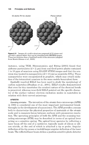
55 atoms Rh fcc cluster Planar defect
x
x
z
z
y
y
Figure 4.13 Images of a stable structure composed of 55 atoms and
one with a planar defect that can be identified with HRTEM images.
These two pictures show a hard-ball model of the structure (adapted
from Marın-Almazo et al., 2005).
instance, using TEM, Hiutsunomiya and Ewing [2003] found that
airborne particulates (d < 2 µm) from coal-fired power plants contained
1 to 10 ppm of uranium using HAADF-STEM images and that the ura-
nium was located in nanoparticles (d < 10 nm) as uraninite (UO ). These
2
nanoparticles were encapsulated in graphite, which may retard oxida-
tion of the tetravalent uranium to the more mobile hexavalent form.
Spatially resolved EELS has been used to study the morphology of
carbon nanotubes [Stephan et al., 2001]. EELS results demonstrated
that even for tiny nanotubes the covalent nature of the chemical bonds
is preserved, whereas near-field EELS pointed out the specific charac-
ter of the surface valence electron excitation modes in nanotubes in
relation with their curved anisotropy.
AFM/STM
Operating principles. The invention of the atomic force microscope (AFM)
in 1982 is considered one of the most important instrumental break-
throughs in the development of nanoscience. The AFM provides a means
both to characterize the physical properties of materials at the atomic
scale and to measure forces between surfaces with piconewton resolu-
tion. The operating principles of both the AFM and the scanning tun-
neling microscope (STM) may be described in terms of an optical lever
acting as a sensitive spring. The optical lever operates by reflecting a
laser beam off the end of a cantilever, typically made of silicon or sili-
con nitride, at the end of which is attached a tip or probe. Angular
deflection of the tip causes a twofold larger angular deflection of the laser
beam. The reflected laser beam strikes a position-sensitive photo-detector