
Page 349 - Failure Analysis Case Studies II
P. 349
334
2.2. Visual inspection
To make a detailed observation of the cracked specimen, specimen D was cut in a direction
perpendicular to the primary crack as depicted in Fig. 2, generating two small sub-specimens, D,
and Dz. With the specimen D, the primary crack was opened to fracture with intention of observing
the crack surface. The crack tip region of the specimen D2 which was not opened was observed by
a microscope. Figure 3(a) and (b) show the inner and outer surfaces of the other specimen DZ.
All other failed tube samples as well as specimen D showed many locally thinned areas at the
inside of the tubes. As observed from Fig. 3(b), which shows the inner surface of the specimen D2,
thinning was fairly localized and occurred irregularly. It should be noted that thinning did not
occur gradually with distance from the burner. From this observation, we can rule out the
possibility of erosion damage by solid particles included in the burner combustion gas as the main
cause of tube thinning. Hence, it may be predicted that local oxidation or local corrosion is the
main causes of thinning.
A thick oxide scale indicated by an arrow in Fig. 3(b) was attached at the locally thinned area.
Similar oxide scales were also observed in a number of local oxidation pits. Most of the oxide
scales contain several cracks formed in random directions. The oxide scale in Fig. 3(b) is enlarged
in Fig. 4. Cracking of the scale must be mainly due to the difference in thermal expansion coefficient
between the oxide scale and the tube metal on which the scale is attached.
Generally at the initial stage of oxidation, an oxide film forms on the metal to prevent further
oxidation. Therefore, when a stabilized oxide film is formed on the surface, the resistance to
oxidation under high temperature conditions is increased. As for the radiant tube of this failure
analysis, the high content (25%) of Cr enables the formation of a Cr203 film that increases
resistance to high temperature oxidation. If this oxide film is removed, the base metal of the tube
will undergo repeated oxidation which results in continuous thickness reduction [2]. When cracking
occurs in the oxide scale, as illustrated in Fig. 4, the crack tip area loses the protective effect of the
oxide film and the base metal beneath the crack tip will be repeatedly oxidised. As a result, a sharp
oxide spike will be gradually formed in the base metal under the oxidation layer where oxide
cracking occurred. Observation of the opened fracture surface of specimen D1 showed an oxide
layer extending to the crack tip.
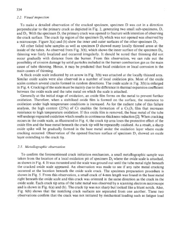
2.3. Metallographic observation
To confirm the forementioned crack initiation mechanism, a small metallographic sample was
taken from the location of a local oxidation pit of specimen D2 where the oxide scale is attached,
as shown in Fig. 4. It was mounted and the scale was ground out until the tube metal right beneath
the cracked oxide scale appeared. An observation was made to see if any tube metal cracking
occurred at the location beneath the oxide scale crack. The specimen preparation procedure is
shown in Fig. 5. From this observation, a small crack of 4-mm length was found in the base metal
right beneath the oxide scale and this crack was oriented in the same direction as the crack in the
oxide scale. Each crack tip area of the tube metal was observed by a scanning electron microscope
and is shown in Fig. 6(a) and (b). The crack tip was not sharp but looked like a blunt notch. Also,
Fig. 6(b) shows that the matching crack surfaces are separated from one another. These two
observations confirm that the crack was not initiated by mechanical loading such as fatigue load