
Page 132 - Fundamentals of Light Microscopy and Electronic Imaging
P. 132
DARK-FIELD MICROSCOPY 115
Paraboloidal Cardioid
(a) (b)
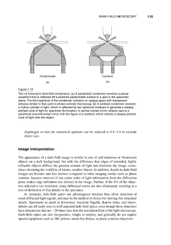
Figure 7-14
Two oil immersion dark-field condensers. (a) A paraboloid condenser receives a planar
wavefront that is reflected off a polished paraboloidal surface to a spot in the specimen
plane. The front aperture of the condenser contains an opaque glass with transparent
annulus similar to that used in phase contrast microscopy. (b) A cardioid condenser receives
a hollow cylinder of light, which is reflected by two spherical surfaces to generate a steeply
pitched cone of light for specimen illumination: A central convex mirror reflects rays to a
peripheral circumferential mirror with the figure of a cardioid, which reflects a steeply pitched
cone of light onto the object.
diaphragm so that the numerical aperture can be reduced to 0.9–1.0 to exclude
direct rays.
Image Interpretation
The appearance of a dark-field image is similar to one of self-luminous or fluorescent
objects on a dark background, but with the difference that edges of extended, highly
refractile objects diffract the greatest amount of light and dominate the image, some-
times obscuring the visibility of fainter, smaller objects. In addition, details in dark-field
images are broader and less distinct compared to other imaging modes such as phase
contrast, because removal of one entire order of light information from the diffraction
plane makes edge definition less distinct in the image. Further, if the NA of the objec-
tive selected is too restricted, many diffracted waves are also eliminated, resulting in a
loss of definition of fine details in the specimen.
In summary, dark-field optics are advantageous because they allow detection of
weak diffracted light signals, and may be the method of choice for viewing fine structural
details. Specimens as small as lysosomes, bacterial flagella, diatom striae, and micro-
tubules are all easily seen in well-adjusted dark-field optics, even though these structures
have dimensions that are 20-times less than the resolution limit of the light microscope.
Dark-field optics are also inexpensive, simple to employ, and generally do not require
special equipment such as DIC prisms, strain-free lenses, or phase contrast objectives.