
Page 130 - Fundamentals of Light Microscopy and Electronic Imaging
P. 130
DARK-FIELD MICROSCOPY 113
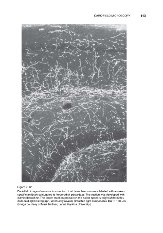
Figure 7-12
Dark-field image of neurons in a section of rat brain. Neurons were labeled with an axon-
specific antibody conjugated to horseradish peroxidase. The section was developed with
diaminobenzidine. The brown reaction product on the axons appears bright white in this
dark-field light micrograph, which only reveals diffracted light components. Bar 100 m.
(Image courtesy of Mark Molliver, Johns Hopkins University.)