
Page 131 - Fundamentals of Light Microscopy and Electronic Imaging
P. 131
114 PHASE CONTRAST MICROSCOPY AND DARK-FIELD MICROSCOPY
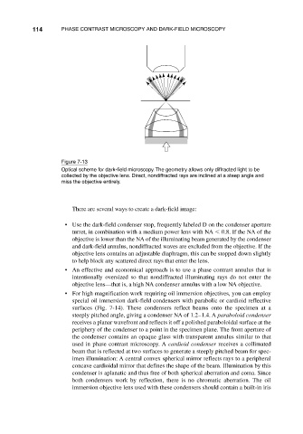
Figure 7-13
Optical scheme for dark-field microscopy. The geometry allows only diffracted light to be
collected by the objective lens. Direct, nondiffracted rays are inclined at a steep angle and
miss the objective entirely.
There are several ways to create a dark-field image:
• Use the dark-field condenser stop, frequently labeled D on the condenser aperture
turret, in combination with a medium power lens with NA 0.8. If the NA of the
objective is lower than the NA of the illuminating beam generated by the condenser
and dark-field annulus, nondiffracted waves are excluded from the objective. If the
objective lens contains an adjustable diaphragm, this can be stopped down slightly
to help block any scattered direct rays that enter the lens.
• An effective and economical approach is to use a phase contrast annulus that is
intentionally oversized so that nondiffracted illuminating rays do not enter the
objective lens—that is, a high NA condenser annulus with a low NA objective.
• For high magnification work requiring oil immersion objectives, you can employ
special oil immersion dark-field condensers with parabolic or cardioid reflective
surfaces (Fig. 7-14). These condensers reflect beams onto the specimen at a
steeply pitched angle, giving a condenser NA of 1.2–1.4. A paraboloid condenser
receives a planar wavefront and reflects it off a polished paraboloidal surface at the
periphery of the condenser to a point in the specimen plane. The front aperture of
the condenser contains an opaque glass with transparent annulus similar to that
used in phase contrast microscopy. A cardioid condenser receives a collimated
beam that is reflected at two surfaces to generate a steeply pitched beam for spec-
imen illumination: A central convex spherical mirror reflects rays to a peripheral
concave cardioidal mirror that defines the shape of the beam. Illumination by this
condenser is aplanatic and thus free of both spherical aberration and coma. Since
both condensers work by reflection, there is no chromatic aberration. The oil
immersion objective lens used with these condensers should contain a built-in iris