Page 217 - Fundamentals of Light Microscopy and Electronic Imaging
P. 217
200      FLUORESCENCE MICROSCOPY


                                   entire text of the exercise before beginning the work. Work in pairs to make the
                                   exercise go smoothly.
                                      In this exercise we will stain tissue culture cells with organelle-specific fluo-
                                   rescent dyes to examine the morphology and abundance of organelle systems and
                                   to compare the performance characteristics of different filter sets. Many, but not
                                   all, of these dyes can be used to stain cells in the living state. Large well-spread
                                   cells, such as COS-7, U2OS, CHO, LLCPK cells, or primary cultures of endothe-
                                   lial cells, if available, are appropriate for this exercise. Prepare representative
                                   sketches of the labeled cells. Keep a record of your observations, including the
                                   time course of the staining response, the stability of fluorescence, and other rele-
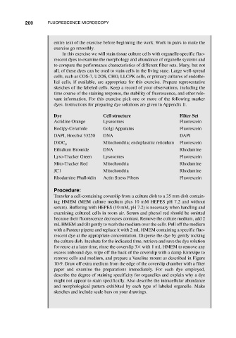
                                   vant information. For this exercise pick one or more of the following marker
                                   dyes. Instructions for preparing dye solutions are given in Appendix II.

                                   Dye                  Cell structure                  Filter Set
                                   Acridine Orange      Lysosomes                       Fluorescein
                                   Bodipy-Ceramide      Golgi Apparatus                 Fluorescein
                                   DAPI, Hoechst 33258  DNA                             DAPI
                                   DiOC 6               Mitochondria; endoplasmic reticulum  Fluorescein
                                   Ethidium Bromide     DNA                             Rhodamine
                                   Lyso-Tracker Green   Lysosomes                       Fluorescein
                                   Mito-Tracker Red     Mitochondria                    Rhodamine
                                   JC1                  Mitochondria                    Rhodamine
                                   Rhodamine Phalloidin  Actin Stress Fibers            Fluorescein

                                   Procedure:
                                   Transfer a cell-containing coverslip from a culture dish to a 35 mm dish contain-
                                   ing HMEM (MEM culture medium plus 10 mM HEPES pH 7.2 and without
                                   serum). Buffering with HEPES (10 mM, pH 7.2) is necessary when handling and
                                   examining cultured cells in room air. Serum and phenol red should be omitted
                                   because their fluorescence decreases contrast. Remove the culture medium, add 2
                                   mL HMEM and tilt gently to wash the medium over the cells. Pull off the medium
                                   with a Pasteur pipette and replace it with 2 mL HMEM containing a specific fluo-
                                   rescent dye at the appropriate concentration. Disperse the dye by gently rocking
                                   the culture dish. Incubate for the indicated time, retrieve and save the dye solution
                                   for reuse at a later time, rinse the coverslip 3  with 1 mL HMEM to remove any
                                   excess unbound dye, wipe off the back of the coverslip with a damp Kimwipe to
                                   remove cells and medium, and prepare a Vaseline mount as described in Figure
                                   10-9. Draw off extra medium from the edge of the coverslip chamber with a filter
                                   paper and examine the preparations immediately. For each dye employed,
                                   describe the degree of staining specificity for organelles and explain why a dye
                                   might not appear to stain specifically. Also describe the intracellular abundance
                                   and morphological pattern exhibited by each type of labeled organelle. Make
                                   sketches and include scale bars on your drawings.

