
Page 214 - Fundamentals of Light Microscopy and Electronic Imaging
P. 214
THE PROBLEM OF BLEED-THROUGH WITH MULTIPLY STAINED SPECIMENS 197
1600
1200
Amplitude 800
400
** * Distance
(a) (b)
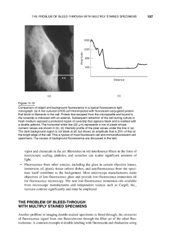
Figure 11-10
Comparison of object and background fluorescence in a typical fluorescence light
micrograph. (a) A live cultured U2OS cell microinjected with fluorescein-conjugated protein
that binds to filaments in the cell. Protein that escaped from the micropipette and bound to
the coverslip is indicated with an asterisk. Subsequent retraction of the cell during culture in
fresh medium exposed a protected region of coverslip that appears black and is marked with
a double asterisk. The horizontal white line (22 m) represents a row of pixels whose
numeric values are shown in (b). (b) Intensity profile of the pixel values under the line in (a).
The dark background region is not black at all, but shows an amplitude that is 25% of that at
the bright edge of the cell. This is typical of most fluorescent cell and immunofluorescent cell
specimens. The causes of background fluorescence are discussed in the text.
vapor and chemicals in the air. Blemishes in old interference filters in the form of
microscopic scaling, pinholes, and scratches can scatter significant amounts of
light.
• Fluorescence from other sources, including the glass in certain objective lenses,
immersion oil, plastic tissue culture dishes, and autofluorescence from the speci-
men itself contribute to the background. Most microscope manufacturers make
objectives of low-fluorescence glass and provide low-fluorescence immersion oil
for fluorescence microscopy. The new low-fluorescence immersion oils available
from microscope manufacturers and independent sources such as Cargill, Inc.,
increase contrast significantly and must be employed.
THE PROBLEM OF BLEED-THROUGH
WITH MULTIPLY STAINED SPECIMENS
Another problem in imaging double-stained specimens is bleed-through, the crossover
of fluorescence signal from one fluorochrome through the filter set of the other fluo-
rochrome. A common example is double labeling with fluorescein and rhodamine using