
Page 245 - Fundamentals of Light Microscopy and Electronic Imaging
P. 245
228 CONFOCAL LASER SCANNING MICROSCOPY
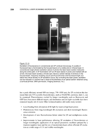
Figure 12-13
Excitation of fluorescence in conventional and 2P confocal microscopy. A cuvette of
fluorescent dye is illuminated by the focused beams of a visible and an infrared laser. In
conventional single-photon excitation (above in photo) the laser beam excites molecules
along the entire path; in 2P illumination with an IR laser (below in photo and marked with an
arrow), the laser beam excites a minute spot. Above a certain density of photons in the
focused beam, frequency doubling occurs and fluorochromes emit fluorescent light, but
below the critical density no 2P excitation occurs. By controlling the laser power, excitation
can be induced in a volume that is close to the thickness of an optical section obtained using
a confocal pinhole. (With permission, Imaging Sciences, Inc.)
has a peak efficiency around 800 nm (range, 750–1050 nm), for 2P excitation this has
meant that only UV-excitable fluorochromes, such as NAD(P)H, serotonin, Indo-1, can
be examined. Fluorochromes excited by longer wavelengths such as fluorescein and
GFP have been more difficult targets, and rhodamine and red light–excitable dyes have
remained largely out of reach. Other technical matters still under study include:
• Local heating from absorption of IR light by water at high laser power.
• Phototoxicity from long-wavelength IR excitation and short-wavelength fluores-
cence emission.
• Development of new fluorochromes better suited for 2P and multiphoton excita-
tion.
• Improvements in laser performance allowing 2P excitation of fluorochromes at
longer wavelengths; application of an optical parametric oscillator pumped by a
titanium:sapphire laser and the use of neodymium lasers allow efficient 2P excita-
tion at a wide range of UV and visible wavelengths.