
Page 246 - Fundamentals of Light Microscopy and Electronic Imaging
P. 246
CONFOCAL IMAGING WITH A SPINNING NIPKOW DISK 229
Excellent examples of the application of 2-photon imaging in neuroscience applications
are given by Denk and others in Yuste et al. (2000).
CONFOCAL IMAGING WITH A SPINNING NIPKOW DISK
Instead of illuminating the object by raster scanning using a single spot, it is possible to
scan the specimen with thousands of points simultaneously using a spinning Nipkow
disk (Fig. 12-14). A Nipkow disk contains thousands of minute pinholes arranged in
rows of outwardly spiraling tracks. The arrangement and spacing of the pinholes is such
that every point in the specimen receives the same amount of illumination from the
rotating disk. There are substantial advantages inherent to this design:
• The returned fluorescent light can generate a real confocal image that can be seen
by the eye or recorded on a camera, so no PMT-based imaging system is required.
Spread laser
beam
Microlens array
Dichroic mirror
Lens
CCD camera
Pinhole array
Rotation
Objective
Sample
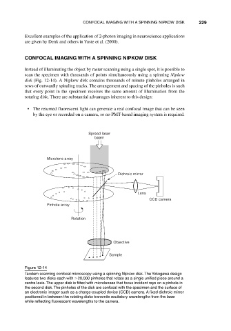
Figure 12-14
Tandem scanning confocal microscopy using a spinning Nipkow disk. The Yokogawa design
features two disks each with 20,000 pinholes that rotate as a single unified piece around a
central axis. The upper disk is fitted with microlenses that focus incident rays on a pinhole in
the second disk. The pinholes of the disk are confocal with the specimen and the surface of
an electronic imager such as a charge-coupled device (CCD) camera. A fixed dichroic mirror
positioned in between the rotating disks transmits excitatory wavelengths from the laser
while reflecting fluorescent wavelengths to the camera.