
Page 251 - Fundamentals of Light Microscopy and Electronic Imaging
P. 251
234 VIDEO MICROSCOPY
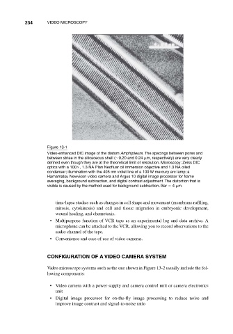
Figure 13-1
Video-enhanced DIC image of the diatom Amphipleura. The spacings between pores and
between striae in the silicaceous shell ( 0.20 and 0.24 m, respectively) are very clearly
defined even though they are at the theoretical limit of resolution. Microscopy: Zeiss DIC
optics with a 100 , 1.3 NA Plan Neofluar oil immersion objective and 1.3 NA oiled
condenser; illumination with the 405 nm violet line of a 100 W mercury arc lamp; a
Hamamatsu Newvicon video camera and Argus 10 digital image processor for frame
averaging, background subtraction, and digital contrast adjustment. The distortion that is
visible is caused by the method used for background subtraction. Bar 4 m.
time-lapse studies such as changes in cell shape and movement (membrane ruffling,
mitosis, cytokinesis) and cell and tissue migration in embryonic development,
wound healing, and chemotaxis.
• Multipurpose function of VCR tape as an experimental log and data archive. A
microphone can be attached to the VCR, allowing you to record observations to the
audio channel of the tape.
• Convenience and ease of use of video cameras.
CONFIGURATION OF A VIDEO CAMERA SYSTEM
Video microscope systems such as the one shown in Figure 13-2 usually include the fol-
lowing components:
• Video camera with a power supply and camera control unit or camera electronics
unit
• Digital image processor for on-the-fly image processing to reduce noise and
improve image contrast and signal-to-noise ratio