
Page 48 - Handbook of Adhesion Promoters
P. 48
2.13 Cellular adhesion 41
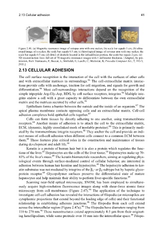
Figure 2.46. (a) Magnetic resonance image of octopus arm with one sucker, the scale bar equals 1 cm; (b) ultra-
sound image of a sucker, the scale bar equals 0.5 cm; (c) histological image of octopus arm with one sucker, the
scale bar equals 0.5 cm; (d) detail of denticle located in the infundibular portion, the scale bar equals 2 μm; (e)
3D reconstruction from full set of 70 magnetic resonance images with 1 millimeter thickness. [Adapted, by per-
mission, from Tramacere, F; Beccai, L; Sinibaldi, E; Laschi, C; Mazzolai, B, Procedia Computer Sci., 7, 192-3,
2011.]
2.13 CELLULAR ADHESION
The cell surface recognition is the interaction of the cell with the surfaces of other cells
86
and with extracellular matrices in surroundings. The cell-extracellular matrix interac-
tions provide cells with anchorage, traction for cell migration, and signals for growth and
86
differentiation. Most cell-surroundings interactions depend on the recognition of the
86
simple tripeptide Arg-Gly-Asp, RDG, by cell surface receptors, integrins. Multiple inte-
grins endow a cell with a great capacity to differentiate between the own extracellular
86
matrix and the matrices secreted by other cells.
87
Epithelium forms a barrier between the outside and the inside of an organism. The
apical plasma membrane contacts opposing cells and an extracellular matrix. Cell-cell
87
adhesion complexes hold epithelial cells together.
Cells can form tissues by directly adhering to one another, using transmembrane
88
receptors. Another means of adhesion is to attach the cell to the extracellular matrix,
88
ECM, (dynamic, highly crosslinked mesh of insoluble proteins). This is primarily medi-
88
ated by the transmembrane integrin receptors. They anchor the cell and provide an indi-
rect means of cell-cell adhesion when different cells connect to a common ECM between
88
them. These features play critical roles in the construction and maintenance of tissues
88
during development and adult life.
Keratin is a protein of human hair but it is also a protein which regulates the func-
89
89
tions of the liver. Hepatocytes are the cells of the liver tissue. Hepatocytes make up 70-
89
85% of the liver's mass. The keratin biomaterials researchers, aiming at regulating phys-
iological events through surface-mediated control of cellular behavior, are interested in
89
adhesion between human hair keratins and hypatocytes. The hepatocyte adhesion to ker-
atin substrates was not mediated by integrins of the β - or β -subtype but by hepatic glyco-
1
2
89
protein receptor. Glycopolymer surfaces preserve the differentiated state of mature
89
hepatocytes and help maintain their ability to perform liver-specific functions.
Scanning near-field optical microscopy, SNOM, has been employed to simultane-
ously acquire high-resolution fluorescence images along with shear-force atomic force
90
microscopy from cell membranes (Figure 2.47). The application of the technique to
investigate cell-cell adhesion has revealed the interactions of filopodia (or microspikes are
cytoplasmic projections that extend beyond the leading edge of cells) and their functional
90
relationship in establishing adherens junctions. The filopodia from each cell extend
90
across the intercellular region (Figure 2.47a). The filopodia have diameters ranging from
90
110 to 270 nm. These nanostructures extend approximately 4-5 μm from their originat-
90
ing lamellipodium, while some protrude over 10 mm into the intercellular space. Filopo-