
Page 221 - Handbook of Biomechatronics
P. 221
218 Georgios A. Bertos and Evangelos G. Papadopoulos
OPRA ILP OPL
Fixture
Abutment
Abutment screw
1
2
Skin 3
Bone
4
(A) (C) 5
2 3
4
1
5
6
7 8
(B) (D) (E) (F) (G)
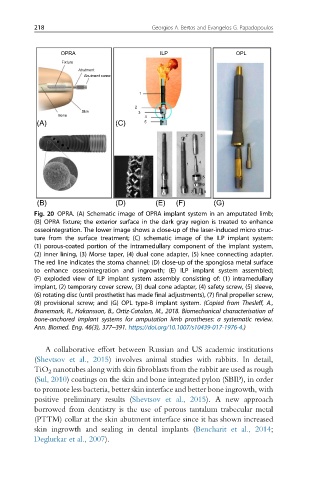
Fig. 20 OPRA. (A) Schematic image of OPRA implant system in an amputated limb;
(B) OPRA fixture; the exterior surface in the dark gray region is treated to enhance
osseointegration. The lower image shows a close-up of the laser-induced micro struc-
ture from the surface treatment; (C) schematic image of the ILP implant system:
(1) porous-coated portion of the intramedullary component of the implant system,
(2) inner lining, (3) Morse taper, (4) dual cone adapter, (5) knee connecting adapter.
The red line indicates the stoma channel; (D) close-up of the spongiosa metal surface
to enhance osseointegration and ingrowth; (E) ILP implant system assembled;
(F) exploded view of ILP implant system assembly consisting of: (1) intramedullary
implant, (2) temporary cover screw, (3) dual cone adapter, (4) safety screw, (5) sleeve,
(6) rotating disc (until prosthetist has made final adjustments), (7) final propeller screw,
(8) provisional screw; and (G) OPL type-B implant system. (Copied from Thesleff, A.,
Branemark, R., Hakansson, B., Ortiz-Catalan, M., 2018. Biomechanical characterisation of
bone-anchored implant systems for amputation limb prostheses: a systematic review.
Ann. Biomed. Eng. 46(3), 377–391. https://doi.org/10.1007/s10439-017-1976-4.)
A collaborative effort between Russian and US academic institutions
(Shevtsov et al., 2015) involves animal studies with rabbits. In detail,
TiO 2 nanotubes along with skin fibroblasts from the rabbit are used as rough
(Sul, 2010) coatings on the skin and bone integrated pylon (SBIP), in order
to promote less bacteria, better skin interface and better bone ingrowth, with
positive preliminary results (Shevtsov et al., 2015). A new approach
borrowed from dentistry is the use of porous tantalum trabecular metal
(PTTM) collar at the skin abutment interface since it has shown increased
skin ingrowth and sealing in dental implants (Bencharit et al., 2014;
Deglurkar et al., 2007).