
Page 60 - Handbook of Properties of Textile and Technical Fibres
P. 60
Testing and characterization of fibers 41
2.3.5.3 Apparent crystallite size
Profile analysis of equatorial scans, shown in Fig. 2.13(a), can be used to obtain an
apparent crystallite size in the normal direction of (hkl) layers using the following
expression (Wilchinsky, 1959):
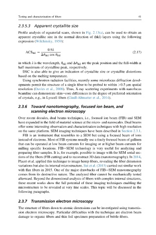
0:9l
ACS hkl ¼ (2.17)
Dq hkl cos q hkl
in which l is the wavelength, q hkl and Dq hkl are the peak position and the full-width at
half-maximum of crystalline peak, respectively.
DSC is also able to give an indication of crystallite size or crystallite distortions
based on the melting temperature.
Using synchrotron radiation facilities, recently some microfocus diffraction devel-
opments permit the structure of a single fiber to be probed to within >0.5 mm spatial
resolution (Davies et al., 2008). Thus, X-ray scattering experiments with nanofocus
beamline can demonstrate skinecore differences in the degree of preferred orientation
of crystals, e.g., in Lyocell fibers (Gindl-Altmutter et al., 2014).
2.3.6 Toward nanotomography, focused ion beam, and
scanning electron microscopy
Over recent decades, dual beams techniques, i.e., focused ion beam (FIB) and SEM
have expanded in the field of material science at the micro- and nanoscales. Dual beams
offer some interesting observation and characterization techniques with high resolution
on the same platform. SEM imaging techniques have been described in Section 2.3.1.
FIB is an instrument that resembles to a SEM but using a focused beam of ions
instead of electrons. Most of FIB systems usually use a finely focused beam of gallium
that can be operated at low beam currents for imaging or at higher beam currents for
milling specific locations. FIBeSEM technology is very useful for analyzing and
preparing fiber samples. It is, for example, possible to image with the SEM serial sec-
tions of the fibers (FIB cutting) and to reconstruct 3D data (nanotomography). In 2014,
Placet et al. applied this technique to image hemp fibers, revealing the fiber dimension
variations but also its internal microstructure. Sui et al. (2015) carried out similar work
with flax fibers in 2015. One of the major drawbacks of FIBeSEM nanotomography
comes from its destructive nature. The analyzed fiber cannot be mechanically tested
afterward. Beyond the dimensional analysis of fibers with complex internal structures,
these recent works show the full potential of these imaging techniques enabling the
microstructure to be revealed at very fine scales. This topic will be discussed in the
following paragraphs.
2.3.7 Transmission electron microscopy
The structure of fibers down to atomic dimensions can be investigated using transmis-
sion electron microscopy. Particular difficulties with the technique are electron beam
damage to organic fibers and thin foil specimen preparation of brittle fibers.