
Page 205 - Introduction to Colloid and Surface Chemistry
P. 205
194 Charged interfaces
-6
-75
-4
-50
(b)
~o
1 -25
£ .
o
a. o
E
o
10
Q.
O
+25-
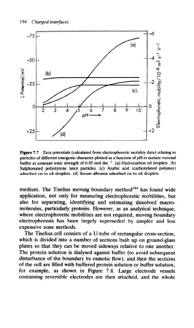
Figure 7.7 Zeta potentials (calculated from electrophoretic mobility data) relating to
particles of different ionogenic character plotted as a function of pH in acetate-veronal
3
buffer at constant ionic strength of 0.05 mol dm~ . (a) Hydrocarbon oil droplets, (b)
Sulphonated polystyrene latex particles, (c) Arabic acid (carboxylated polymer)
adsorbed on to oil droplets, (d) Serum albumin adsorbed on to oil droplets
medium. The Tiselius moving boundary method 184 has found wide
application, not only for measuring electrophoretic mobilities, but
also for separating, identifying and estimating dissolved macro-
molecules, particularly proteins. However, as an analytical technique,
where electrophoretic mobilities are not required, moving boundary
electrophoresis has been largely superseded by simpler and less
expensive zone methods.
The Tiselius cell consists of a U-tube of rectangular cross-section,
which is divided into a number of sections built up on ground-glass
plates so that they can be moved sideways relative to one another.
The protein solution is dialysed against buffer (to avoid subsequent
disturbance of the boundary by osmotic flow), and then the sections
of the cell are filled with buffered protein solution or buffer solution,
for example, as shown in Figure 7.8. Large electrode vessels
containing reversible electrodes are then attached, and the whole