
Page 68 - Mechanics of Asphalt Microstructure and Micromechanics
P. 68
Microstructure Characterization 61
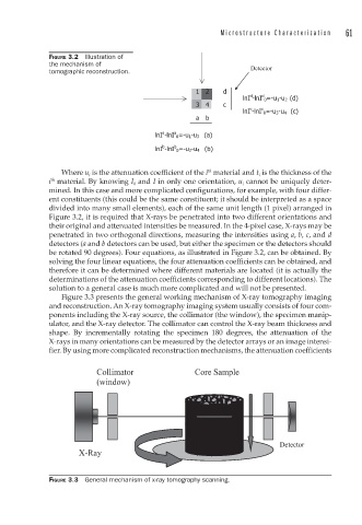
FIGURE 3.2 Illustration of
the mechanism of
tomographic reconstruction.
th
Where u i is the attenuation coefficient of the i material and t i is the thickness of the
i material. By knowing I 0 and I in only one orientation, u i cannot be uniquely deter-
th
mined. In this case and more complicated configurations, for example, with four differ-
ent constituents (this could be the same constituent; it should be interpreted as a space
divided into many small elements), each of the same unit length (1 pixel) arranged in
Figure 3.2, it is required that X-rays be penetrated into two different orientations and
their original and attenuated intensities be measured. In the 4-pixel case, X-rays may be
penetrated in two orthogonal directions, measuring the intensities using a, b, c, and d
detectors (a and b detectors can be used, but either the specimen or the detectors should
be rotated 90 degrees). Four equations, as illustrated in Figure 3.2, can be obtained. By
solving the four linear equations, the four attenuation coefficients can be obtained, and
therefore it can be determined where different materials are located (it is actually the
determinations of the attenuation coefficients corresponding to different locations). The
solution to a general case is much more complicated and will not be presented.
Figure 3.3 presents the general working mechanism of X-ray tomography imaging
and reconstruction. An X-ray tomography imaging system usually consists of four com-
ponents including the X-ray source, the collimator (the window), the specimen manip-
ulator, and the X-ray detector. The collimator can control the X-ray beam thickness and
shape. By incrementally rotating the specimen 180 degrees, the attenuation of the
X-rays in many orientations can be measured by the detector arrays or an image intensi-
fier. By using more complicated reconstruction mechanisms, the attenuation coefficients
FIGURE 3.3 General mechanism of x-ray tomography scanning.