
Page 125 -
P. 125
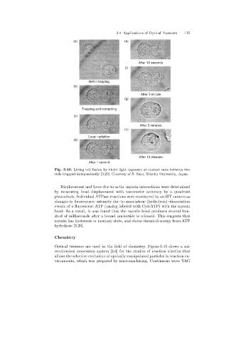
3.4 Applications of Optical Tweezers  115
                                     (a)                       (e)


                                                                     After 10 seconds
                                                               (f)


                                             Befor trapping
                                     (b)

                                                                       After 1 minute
                                                               (g)

                                         Trapping and contacting
                                     (c)

                                                                      After 5 minutes
                                                               (h)
                                             Laser radiation
                                     (d)


                                                                      After 15 minutes
                                             After 1 second
                            Fig. 3.40. Living cell fusion by violet light exposure at contact area between two
                            cells trapped independently [3.20]. Courtesy of S. Sato, Tohoku University, Japan


                               Displacement and force due to actin–myosin interactions were determined
                            by measuringbead displacement with nanometer accuracy by a quadrant
                            photodiode. Individual ATPase reactions were monitored by an SIT camera as
                            changes in fluorescence intensity due to association–(hydrolysis)–dissociation
                            events of a fluorescent ATP (analoglabeled with Cy3-ATP) with the myosin
                            head. As a result, it was found that the myosin head produces several hun-
                            dred of milliseconds after a bound nucleotide is released. This suggests that
                            myosin has hysteresis or memory state, and stores chemical energy from ATP
                            hydrolysis [3.26].

                            Chemistry

                            Optical tweezers are used in the field of chemistry. Figure 3.43 shows a mi-
                            crochemical conversion system [3.6] for the studies of reaction kinetics that
                            allows the selective excitation of optically manipulated particles in reaction en-
                            vironments, which was prepared by micromachining. Continuous wave YAG