
Page 433 - Book Hosokawa Nanoparticle Technology Handbook
P. 433
7.3 SAFETY OF NANOPARTICLES FUNDAMENTALS
5
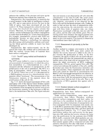
enhances the stability of the structure and raises up the that cyto-toxicity is not observed for 10 cells when the
fluorescent intensity than without the coated one. concentration is less than 0.5 M. This result means
Nanoparticles, thus manufactured, as materials for that this concentration is the threshold of the cell toxi-
a novel memory in the field of intelligence technol- city. Likewise, cyto-toxicity is not observed for the
ogy (IT), and as super-micro devices for laser in the HeLa cells and for the human primary cells. Further, in
field of optics, have been developed all over the order to find out how the sizes of nanoparticles influ-
world [1–5]. These nanoparticles cannot be dissolved ence the cyto-toxicity, the cyto-toxicities were evalu-
into water, but dissolved into organic solvents like ated with three kinds of quantum dots; one whose
toluene. Therefore, for biological and medical appli- fluorescence wavelength is 640 nm, red, one with 570
cations, various technologies for surface-conjugations nm, yellow, and the other with 520nm, green. The fol-
to make them hydrophilic [6, 7] have been developed. lowing results were obtained. The largest quantum dots
For example, nanoparticles covered with TOPO are whose fluorescence wavelength is 640 nm show a ten-
hydrophobic because an alkyl group on them is dency to give cyto-toxicity. Cyto-toxicity is observed at
hydrophobic. Therefore, a technology for replacing concentrations more than 1 M [11].
this alkyl group with hydrophilic carbonic acid (mak-
ing the whole particles soluble in water) has been 7.3.3.2 Measurement of cyto-toxicity by the flow
developed [6]. cytometry
Nanoparticles, thus surface-treated, can be dis-
solved into water, like sodium salt or potassium salt. Another method to evaluate cyto-toxicity is the flow
With this method, various kinds of materials have cytometry [12]. The MTT assay alone cannot tell
been surface-conjugated. whether the toxicity observed is lethal to the cells or
just restrains the prolification of them. In the flow
7.3.3.1 The MTT assay method and thresholds for cytometry, the nuclei of dead cells are dyed with pro-
cyto-toxicity pidium iodide (PI) after the nanoparticles are taken in
and the ratios of the dead cells are measured. Fig.7.3.6
The MTT assay method is a way to evaluate the haz-
ard assessment of nanoparticles, in which the activa- shows the lethal cyto-toxicity of MUA conjugated
tion metabolism in a mitochondrium in a cell is nanoparticles (520 nm, green) against Vero cells. The
measured and the influence of nanoparticles on the vertical axis indicates the numbers of the cells, and the
prolification of the cell is qualitified. The MTT is a horizontal axes show the fluorescence intensities and
kind of tetrazolium, whose molecular formula is the cyto-toxicities. These experiments also show that
C H BrN S. Taken into a cell, it is decomposed by dead cells cannot be observed at concentrations less
16
18
5
a dehydrogenase enzyme in a mitochondrium into a than 0.5 M even though nanoparticles are taken in, as
pigment called ‘hormazan’. The measurement of the was shown in the MTT assay. However, at concentra-
fluorescence intensity of the pigment shows the tions more than 2 M, the nanoparticles taken in cause
prolification of the cell [8–10]. damage to more than the half of the cells. That is, the
Fig. 7.3.5. shows the hazard assessment of Vero cells cyto-toxicity of MUA quantum dots against cells is
and kidney cells of the African Green Monkey against lethal [11].
CdSe/ZnS nanoparticles. The horizontal axis shows the
concentrations of the nanoparticles and the vertical axis 7.3.3.3 Relations between surface-conjugations of
the fluorescence intensity of the hormazan at 460 nm, nanoparticles and their safety
that is, the metabolism of the cells. The figure indicates Nanoparticles have been surface-conjugated for
applications for various uses.
Some surface-conjugations cause more grave
cyto-toxicity than others. Therefore, relations
between surface-conjugations and their safety for
cells have to be considered. In order to find out the
relations, the safety evaluations of nanoparticles sur-
face-conjugated with two materials were made; one
is with MUA (quantum dots-COOH) and the other is
with glycerol (quantum dots-OH), and their purified
and unpurified particles. Fig. 7.3.7 shows that the
purification reduces the cyto-toxicity for the quan-
tum dots-OH, and that the toxicity remains the same
after the purification for the quantum dots-COOH.
MUA itself, a material with which particles are con-
jugated, has cyto-toxicity. This experiment shows
that toxicity against cells is connected not only with
Figure 7.3.5 particles themselves but also with kinds of surface-
Cyto-toxicity of the quantum dots by the MTT assay. conjugations and degrees of purification.
407