
Page 428 - Book Hosokawa Nanoparticle Technology Handbook
P. 428
FUNDAMENTALS CH. 7 ENVIRONMENTAL AND SAFETY ISSUES WITH NANOPARTICLES
7.3.2.1 Exposure routes and uptake of nanoparticles
(1) Exposure routes for nanoparticles Nasal airway Nasopharyngeal region
Nanoparticles can either be deliberately introduced Pharynx
into the body for medical purposes (drug delivery sys-
tems) or absorbed involuntarily from the environment Larynx
(inhalation of nanoparticle-containing dust in the air). Trachea
A distinction should also be drawn between nanoparti- Tracheobronchial
cles manufactured for industrial application and those Bronchi region
unintentionally generated and released in the environ-
ment, such as welding fumes or diesel exhaust parti-
cles (DEP). In the fields of environmental science and
toxicology, numerous studies on the potential health
hazards caused by ultrafine particles have been con-
ducted. Practically, there are several definitions of Alveolar region
nanoparticles or ultrafine particles, however, findings
regarding biological effects of the ultrafine particles
are useful as a starting point for estimating the effects Alveolar ducts
of nanoparticles on human health.
Human and animals contact with nanoparticles
through various routes: nanoparticles can be inhaled in Alveolar sacs
the air, swallowed in the water, ingested in food, and
absorbed via the skin in cosmetics. For successful risk
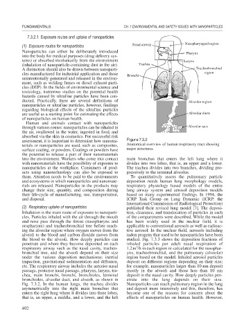
assessment, it is important to determine how nanoma- Figure 7.3.2
terials or nanoparticles are used, such as composites, Anatomical overview of human respiratory tract showing
surface coating, or powders. Coatings or powders have major structures.
the potential to release a part of their nanomaterials
into the environment. Workers who come into contact main bronchus that enters the left lung where it
with nanomaterials have the possibility of exposure to divides into two lobes, that is, an upper and a lower.
nanoparticles at the workplace. Consumers of prod- The trachea divides into two branches, dividing pro-
ucts using nanotechnology can also be exposed to gressively to the terminal alveolus.
them. Attention needs to be paid to the environments To quantitatively assess the pulmonary particle
and ecosystems in which nanoparticles and nanomate- deposition needs human lung morphology models,
rials are released. Nanoparticles in the products may respiratory physiology based models of the entire
change their size, quantity, and composition during lung airway system and aerosol deposition models
their life-cycle of manufacturing, use, transportation, based on many experimental findings. In 1994, the
and disposal. ICRP Task Group on Lung Dynamic (ICRP: the
International Commission of Radiological Protection)
(2) Respiratory uptake of nanoparticles published their revised lung model [7]. The deposi-
Inhalation is the main route of exposure to nanoparti- tion, clearance, and translocation of particles in each
cles. Particles inhaled with the air through the mouth of the compartments were described. While the model
and nose pass through the throat (nasopharynx and has been widely used in the nuclear field, it is
oropharynx) and tracheobronchial tree before reach- applicable to conventional aerosols as well as radioac-
ing the alveolar region where oxygen moves from the tive aerosol. In the nuclear field, aerosols including
alveoli to the blood and carbon dioxide moves from radon progeny that used to be nanoparticles have been
the blood to the alveoli. How deeply particles can studied. Fig. 7.3.3 shows the deposition fractions of
penetrate and where they become deposited on each inhaled particles per adult nasal respiration of
3
respiratory airway such as the nasal cavity, tracheo- 1.2m /h in each region re-calculated for the nasophar-
bronchial tree, and the alveoli depend on their size ynx, tracheobronchial, and the pulmonary (alveolar)
under the various deposition mechanisms: inertial region based on the model. Inhaled aerosol particles
impaction, gravitational sedimentation and diffusion, deposit on different regions depending on their size;
etc. The respiratory airway includes the anterior nasal for example, nanoparticles larger than 10 nm deposit
passage, posterior nasal passage, pharynx, larynx, tra- mostly in the alveoli and those less than 10 nm
chea, main bronchi, bronchi, bronchioles, terminal deposit in the nasal cavity. How deeply particles pen-
bronchioles, alveolar duct, and alveoli, as shown in etrate into the lung depends on their size.
Fig. 7.3.2. In the human lungs, the trachea divides Nanoparticles can reach pulmonary region in the lung
asymmetrically into the right main bronchus that and deposit more intensively and this, therefore, has
enters the right lung where it divides into three lobes, become one of the reasons for concern about the
that is, an upper, a middle, and a lower, and the left effects of nanoparticles on human health. However,
402