
Page 242 - Optofluidics Fundamentals, Devices, and Applications
P. 242
Bio-Inspir ed Fluidic Lenses for Imaging and Integrated Optics 217
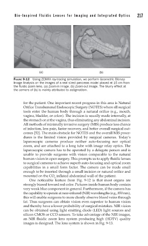
FIGURE 9-12 Using ZEMAX ray-tracing simulation, we perform Geometric Bitmap
Image Analysis on the images of a real sized pancreas model placed at 15 cm from
the fl uidic zoom lens. (a) Zoom-in image. (b) Zoom-out image. The blurry effect at
the corners of (b) is mainly attributed to astigmatism.
for the patient. One important recent progress in this area is Natural
Orifice Translumenal Endoscopic Surgery (NOTES) where all surgical
tools enter the human body through a natural orifice (e.g., mouth,
vagina, bladder, or colon). The incision is usually made internally, at
the stomach or at the vagina, thus eliminating any abdominal incision.
All methods of minimally invasive surgery (MIS) produce less chance
of infection, less pain, faster recovery, and better overall surgical out-
comes [52]. The main obstacle for NOTES and the overall MIS proce-
dures is the limited vision provided by surgical cameras. Today’s
laparoscopic cameras produce neither auto-focusing nor optical
zoom, and are attached to a long tube with image relay optics. The
laparoscopic camera has to be operated by a delegate person and is
unable to provide surgeons with vision comparable to the natural
human vision in open surgery. This prompts us to apply fluidic lenses
to surgical cameras to achieve superb auto-focusing and optical zoom
capabilities in a small form factor. The camera can be made small
enough to be inserted through a small incision or natural orifice and
mounted on the CO inflated abdominal wall of the patient.
2
One noticeable feature from Fig. 9-12 is that most organs are
strongly biased toward red color. Pictures inside human body contain
very weak blue component in general. Furthermore, if the camera has
the capability to operate at near-infrared (NIR) wavelengths (e.g., 850 nm),
this will enable surgeons to more clearly observe blood veins through
fat. Thus surgeons can obtain vision even superior to human vision
and thereby have a lower probability of surgical mistakes. NIR vision
can be obtained using light emitting diode (LED) light sources and
silicon CMOS or CCD sensors. To take advantage of the NIR images,
an NIR fluidic zoom lens system producing high (HDTV) quality
images is designed. The lens system is shown in Fig. 9-13.