
Page 246 - Optofluidics Fundamentals, Devices, and Applications
P. 246
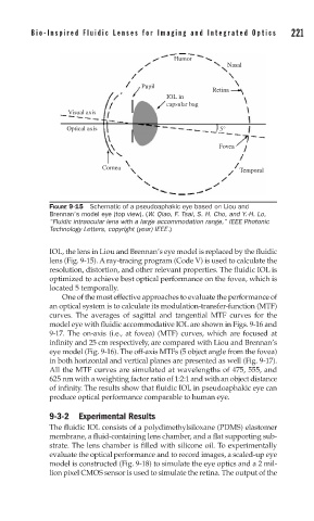
Bio-Inspir ed Fluidic Lenses for Imaging and Integrated Optics   221


                                               Humor
                                                            Nasal

                                      Pupil
                                                        Retina
                                             IOL in
                                             capsular bag
                    Visual axis

                    Optical axis                          5°

                                                          Fovea

                            Cornea
                                                               Temporal


               FIGURE 9-15  Schematic of a pseudoaphakic eye based on Liou and
               Brennan’s model eye (top view). (W. Qiao, F. Tsai, S. H. Cho, and Y.-H. Lo,
               “Fluidic intraocular lens with a large accommodation range,” IEEE Photonic
               Technology Letters, copyright (year) IEEE.)

               IOL, the lens in Liou and Brennan’s eye model is replaced by the fluidic
               lens (Fig. 9-15). A ray-tracing program (Code V) is used to calculate the
               resolution, distortion, and other relevant properties. The fluidic IOL is
               optimized to achieve best optical performance on the fovea, which is
               located 5 temporally.
                  One of the most effective approaches to evaluate the performance of
               an optical system is to calculate its modulation-transfer-function (MTF)
               curves. The averages of sagittal and tangential MTF curves for the
               model eye with fluidic accommodative IOL are shown in Figs. 9-16 and
               9-17. The on-axis (i.e., at fovea) (MTF) curves, which are focused at
               infinity and 25 cm respectively, are compared with Liou and Brennan’s
               eye model (Fig. 9-16). The off-axis MTFs (5 object angle from the fovea)
               in both horizontal and vertical planes are presented as well (Fig. 9-17).
               All the MTF curves are simulated at wavelengths of 475, 555, and
               625 nm with a weighting factor ratio of 1:2:1 and with an object distance
               of infinity. The results show that fluidic IOL in pseudoaphakic eye can
               produce optical performance comparable to human eye.

               9-3-2 Experimental Results
               The fluidic IOL consists of a polydimethylsiloxane (PDMS) elastomer
               membrane, a fluid-containing lens chamber, and a flat supporting sub-
               strate. The lens chamber is filled with silicone oil. To experimentally
               evaluate the optical performance and to record images, a scaled-up eye
               model is constructed (Fig. 9-18) to simulate the eye optics and a 2 mil-
               lion pixel CMOS sensor is used to simulate the retina. The output of the