
Page 279 - Organic Electronics in Sensors and Biotechnology
P. 279
256 Cha pte r S i x
Depending on the nature of the optical filtering employed, the photo-
diode detects the intensity of either the transmitted light from the
OLED or the emitted light from the biolabel; either way, it is possible
to deduce the concentration of the biolabel (and hence the analyte).
Importantly, by splitting the sample stream, it is possible to perform
multiple immunoassays in parallel, which can be monitored using
separate LED/photodetector pairs. In an alternative configuration,
the need for the OLED light source can be eliminated altogether by
using a chemiluminescent assay that generates its own emission. 98
Chemiluminescence (CL) reactions typically involve the formation of
a metastable reaction intermediate in an electronically excited state,
which subsequently relaxes to the ground state with the emission of
a photon. CL is particularly attractive for portable microfluidic assays,
because the CL reaction acts as an internal light source, thereby low-
ering instrumental requirements and significantly reducing power
consumption and background interference compared to fluorescence
assays.
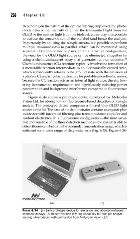
Figure 6.34a shows a prototype device developed by Molecular
Vision Ltd. for absorption- or fluorescence-based detection of a single
analyte. The prototype device comprises a filtered blue OLED light
source in the lid. The base of the demonstrator contains an organic pho-
todetector with integrated filtering plus transimpedance amplifier and
readout electronics. In a fluorescence configuration––the most sensi-
tive and versatile of the three detection methods––the system is able to
detect fluorescent beads in the picomolar concentration range, which is
sufficient for a wide range of diagnostic tests (Fig. 6.35). Figure 6.34b
(a) (b)
FIGURE 6.34 (a) Early prototype device for emission- and absorption-based
chemical assays. (b) Recent version offering capability for multiple-analyte
testing. (Reproduced with permission from Molecular Vision Ltd.)