
Page 287 - Radiochemistry and nuclear chemistry
P. 287
Uses of Radioactive Tracers 271
9.5.3. Emission computer tomography (ECT) and diagnosis
The rate of incorporation and discharge of radioactively labeled substances in the body
provides a measure of the metabolism of healthy and of sick tissues. On medical patients
this information is obtaineA by external measurements referred to as radioisotope scanning
(RIS). Such scanning can yield information about a medical disorder much before it is
observed by other means. Since the amount of radioactive tracer is very small, this
technique is referred to as non-invasive. In hospitals the department of nuclear medicine is
normally responsible for these investigations.
(a) Simple scanners
Simple scanners are designed either with one or several direction sensitive (focusing)
detectors, which are moved around or above the patient in a pattern; Figure 9.13.B and C.
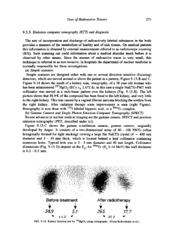
Figure 9.14 shows the result of a kidney scan, renography, of a 38 year old woman who
has been administered 197HGC12 (EC'), tt/~ 2.672 d); in this case a single NaI(TI)-PMT with
collimator was moved in a recti-linear pattern over the kidneys (Fig. 9.13.B). The left
picture shows that 38.9 % of the compound has been fixed to the left kidney, and very little
to the fight kidney. This was caused by a vaginal fibrous sarcoma blocking the urethra from
the fight kidney. After radiation therapy some improvement is seen (fight Figure).
Renography is now done with 131I labeled hippuric acid, or a 99mTc complex.
(b) Gamma Camera and Single Photon Emission Computer Tomography (SPECT)
Recent advances in nuclear medical imaging are the gamma camera, SPECT and positron
emission tomography (PET, described under (c)).
Figure 9.13.C shows the gamma scintillation camera, gamma camera, originally
developed by Anger. It consists of a two-dimensional array of 40 - 100 PMTs (often
hexagonally formed for tight stacking) viewing a large flat NaI(T1) crystal of -- 400 mm
diameter and 5 - 10 mm thick, which is located behind a lead collimator containing
numerous holes. Typical hole size is 2 - 3 mm diameter and 40 mm length. Collimator
dimensions (Fig. 9.13.D) depend on the E.~; for 99mTc (E.y 0.14 MeV) the wall thickness
is 0.2 - 0.3 mm.
. . . "...." .t'
9 9 .. ; ..: ,: ~, .', , .'," .'..:.,;,. ,.:.,~,,.~ I.;I ;','~':' I ," ,.. : . .., !!; ":,.'.:.,'................ ...... . ." .: .,
:',.
*.
t , "'l;).',
.....
I
|t~t~,,'
.'.
:~
0:'
.....
~
9 '~,:; ~I~,~ I. ,.:.' '; '.: '.', (/i '~ ~ '. :: ~.~". ~' . ','. '.. . '. '..'..,, ....; t :,.,,..' ," .... .:,',,,'.' .'.-., ,: ", ..' .. ": .......... 9 . ' .
i~ a#. ~ , ~', ( ," ,',,:,',l '~',~ I,~,1 ~,.... ...... .':., .... .$.11:, .., '.. v ,.:..,.. ; ..... ".
! ,..., =.'-,ri~';,-~.,'~k:':, ....
-' "" ....
! I
!'
.r;:
,', ..,"
,'1 .; .................
1:
I ' " .,.....
, '....,,
/'
. .s
.' ,1' " ~Jl.,;"i.'. ......
,,. ,.' .....
;,, ,.:;~',:.~:, I.,,:. ,.... .... .~~,., .,I| ...... ....::,,,i~,~.,.,,...:.... ,
~l~,: ,', '. 9 '." : 'o I, ':':,~. ,,, I ~ ' .': '...';.',:, r'.'." ..... ~.': ........ .:
;
~ .... ; ~ ~ ~
~'";;"
....
....
. ~/~tr $ .* . .......... I !,.... ::.,::,.,.,.~__.,,,.., ...... ..'~}~,r.r,~','.".'".',"" ' " .. i
'":': " ' ' .....
'.' .'...'.',".'".~'~l;~,'.~
~"
...
...:.:.
.,.. ....
~,:.~.~..
9 .
'.'.'.",
i I . ' " .. ' "","'" ..~,..'.' " ' , ....,:,~. :1".'.'..'" ";~:,,,,:'t~:'~ r!,~:~:::.:.,
:" ' ' ." ; ': I ': '." : '.'..;.:
, " ", '" '. ; ' . :. :'..:~~:',.7," .. .., ::,,.;..~r~,..,.~',...';~::--.
y, ,..;. . : ',' .... , ....,,~,~q~:.~:. . . . . ,. , . .., 9 ..".:~-.-.:;,,;:
....~:~,~,:,;:
",:...~;:.~:L~!i~.-,'....
9 . .. ,..:...-~,?;~-.';:.' ...;..
'i':. ,', . ' . .... ,~,.
.
";'r,,:~u'r; '
Before treatment After radiotherapy
38.9 5.1 3s 17. 7
"~ 44.0 J" "~ ,57.2 ~"
FIG. 9.14. Kidney function test by X97HgCI2 using scintigraphy. (From Kellershohn et al.)