
Page 291 - Radiochemistry and nuclear chemistry
P. 291
Uses of Radioactive Tracers 275
ALZ FLD
34
t
35 36
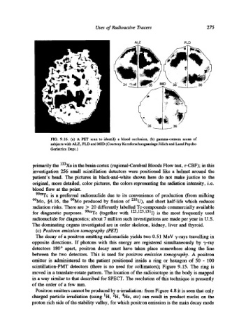
FIG. 9.16. (a) A PET scan to identify a blood occlusion, (b) gamma-camera scans of
subjects with ALZ, FLD and MID (Courtesy Kemforschungsanlage Jiilich and Lurid Psycho
Geriatrics Dept.)
primarily the 133Xe in the brain cortex (regional-Cerebral Bloods Flow test, r-CBF); in this
investigation 256 small scintillation detectors were positioned like a helmet around the
patient's head. The pictures in black-and-white shown here do not make justice to the
original, more detailed, color pictures, the colors representing the radiation intensity, i.e.
blood flow at the point.
99roTe is a preferred radionuclide due to its convenience of production (from milking
99Mo, w the 99Mo produced by fission of 235U), and short half-life which reduces
radiation risks. There are > 20 differently labelled Tc-compounds commercially available
for diagnostic purposes. 99mTc (together with 123'125'1311) is the most frequently used
radionuclide for diagnostics; about 7 million such investigations are made per year in U.S.
The dominating organs investigated are in order skeleton, kidney, liver and thyroid.
(c) Positron emission tomography (PET)
The decay of a positron emitting radionuclide yields two 0.51 MeV v-rays travelling in
opposite directions. If photons with this energy are registered simultaneously by "y-ray
detectors 180 ~ apart, positron decay must have taken place somewhere along the line
between the two detectors. This is used for positron emission tomography. A positron
emitter is administered to the patient positioned inside a ring or hexagon of 50 - 100
scintillation-PMT detectors (there is no need for collimators); Figure 9.15. The ring is
moved in a translate-rotate pattern. The location of the radioisotope in the body is mapped
in a way similar to that described for SPECT. The resolution of this technique is presently
of the order of a few ram.
Positron emitters cannot be produced b~ n-irradiation: from Figure 4.8 it is seen that only
1
.~
4
charged particle irradiation (using H, H, He, etc) can result in product nuclei on the
proton rich side of the stability valley, for which positron emission is the main decay mode