
Page 289 - Radiochemistry and nuclear chemistry
P. 289
Uses of Radioactive Tracers 273
Each picture by the camera is two-dimensional; however, by positioning the camera at
different angles around the patient, 3-dimensional pictures of the organs can be constructed.
This is commonly done by letting the camera rotate around the patient, usually 360 ~ in
increments of a few degrees. Several large cameras at fixed positions or rotating around the
patient can be used for the same purpose. The cameras can also be moved in a direction
parallel to the patient. By using software similar to that used in TCT-scanning,
"radiographic slices" of the patient's organs can be obtained. Such investigations are
referred to as SPECT. The resolution of present commercial SPECT equipment is only 12-
15mm.
With the increasing number of radiopharmaceuticals with specific biological affinities,
gamma camera and SPECT have become important diagnostic tools with numerous clinical
applications, and virtually every organ in the body has been studied. Table 9.4 shows the
most frequently performed imaging investigations; Table 9.5 lists data for radionuclides
applied in medicine, including the amount of radionuclide needed. The radiation doses
received by the patient in diagnostic investigations is usually < 10 mGy per investigation.
The SPECT technique is primarily used for cardiovascular and brain imaging. Cardiac
stress tests, using 2~ or 99mTc labelled radiopharrnaceuticals, amount in the U.S. to
about 2 million a year. Brain tumors can be located by SPECT after intravenous injection
of Na99mTcO4 , as brain tumors have a very high affinity for and slow release of Tc. In
comparison, the uptake of Tc in brain infarcts is low and the release fast, and from healthy
parts of the brain even faster; thus various constrictions to the cerebral blood flow are
easily located. A head scan can be made in 10 minutes and virtually instantaneously
produces an image of the brain.
Mental disorders are diagnosed by SPECT, gamma-camera or PET using various
radiopharmaceuticals, e.g. after the injection of 99mTc-HMPAO (Hexa Methyl Propylene
Amine Oxide) or inhalation of 133Xe. Injecting --- 1000 MBq To-complex into the blood
stream, about 5 % of this compound moves to the brain, passes the membrane of the blood
vessels and enters into the brain tissue, where it decomposes and decays with its 6.0 h half-
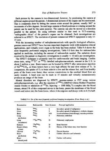
TABLE 9.4. Ten of the most frequently performed imaging investigations (From Sharp et al.)
Examination Radionuclide Static/dynamic Principal application
Bone 99mTc S Secondary spread of malignancy
Liver 99mrc S Secondary spread of malignancy; cirrhotic changes
Brain 99mrc S/D Occult metastases; brain damage; vascular problems
Lung 99mrc S Pulmonary embolism
(perfusion)
Lung 133Xe/99mFc D Pulmonary emphysema
(ventilation)
Kidneys 1231/9~rc D Renal function
Hepato-biliary 99mTc D Patency of the biliary tree; liver function
system
Thyroid 1231/99mrc S Thyroid function
Heart ~~ S Cardiac infarction and ischaemia
(perfusion)
Heart 99mTc D Cardiac wall motion
(blood pool)