
Page 76 - Tandem Techniques
P. 76
Page 57
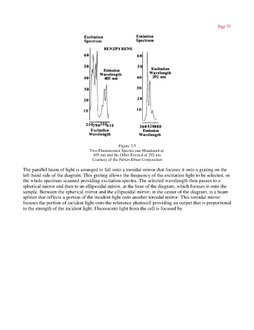
Figure 2.5
Two Fluorescence Spectra one Monitored at
405 nm and the Other Excited at 292 nm
Courtesy of the Perkin Elmer Corporation
The parallel beam of light is arranged to fall onto a toroidal mirror that focuses it onto a grating on the
left-hand side of the diagram. This grating allows the frequency of the excitation light to be selected, or
the whole spectrum scanned providing excitation spectra. The selected wavelength then passes to a
spherical mirror and then to an ellipsoidal mirror, at the base of the diagram, which focuses it onto the
sample. Between the spherical mirror and the ellipsoidal mirror, in the center of the diagram, is a beam
splitter that reflects a portion of the incident light onto another toroidal mirror. This toroidal mirror
focuses the portion of incident light onto the reference photocell providing an output that is proportional
to the strength of the incident light. Fluorescent light from the cell is focused by