
Page 294 - Vibrational Spectroscopic Imaging for Biomedical Applications
P. 294
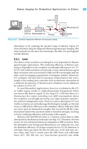
Raman Imaging for Biomedical Applications in Clinics   269


                                      Laser (1)


                                     Filter (3)        Microscope (2)
                                                    DM

                  CCD (5) Spectrograph (4)             Sample
                                       Pinhole
                                                    x, y, z
   FIGURE 9.2 Confocal dispersive Raman microscopic setup.

        direction(s) or by scanning the spectral range of interest. Figure 9.2
        shows the basic setup for dispersive Raman spectroscopic imaging. The
        main elements are the laser, the microscope, the filter, the spectrograph
        and the detector.
        9.2.1 Laser
        The choice of the excitation wavelength is very important for Raman
        spectroscopic applications. The scattering efficiency of Raman scat-
        tering is dependent on the excitation wavelength with a power of 1/λ .
                                                                 4
        So UV and visible excitation will provide a more intense Raman signal
        than excitation with near-infrared (NIR) and IR wavelengths and are
        often used for imaging experiments of inorganic matters. Moreover,
        UV excitation will also lead to resonance enhancement if the wave-
        length of the exciting laser coincides with an electronic absorption of
        a molecule the intensity of Raman-active vibrations can be enhanced
                           4
                      2
        by a factor of 10  to 10 .
            In most biomedical applications, however, excitation in the UV
        or visible region results in a high-fluorescence background which
        can drown the Raman signal. In the deep UV (<250 nm) there is a
        fluorescence-free region extending over 4000 wavenumbers above
        the excitation wavelength providing very high-detection sensitivi-
        ties and low-background noise. However, due to absorption UV and
        visible excitation can photodamage the biological sample, so the best
        choice is NIR (650 to 1400 nm) or IR excitation (1400 to 3000 nm). Both
        are much used for biomedical samples. But toward the IR the water
        absorption band in the spectrum starts to interfere strongly, resulting
        in lower scattering efficiency and heating of the sample.
            Between 650 and 850 nm there is a window where there is little
        absorption by biochemical molecules (see Fig. 9.3). Therefore, the best
        choice for cell and tissue Raman investigation is NIR excitation. Here
        the absorption and background fluorescence are relatively low, and
        the detection efficiency of charge-coupled-device (CCD) detectors is
        still high. Many gas lasers like He-Ne (633 nm), Ar-Kr (647 nm), etc.,
        have been used, but in recent years the NIR diode and solid-state
        lasers (671, 785, 830 nm, etc.) have become powerful and stable