
Page 295 - Vibrational Spectroscopic Imaging for Biomedical Applications
P. 295
270 Cha pte r Ni ne
1.2
Fat Water
1 Hemoglobin
Absorption (a.u.) 0.6
0.8
0.4
0.2
0
450 550 650 750 850 950 1050
λ (nm)
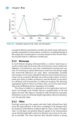
FIGURE 9.3 Absorption spectra of fat, water, and hemoglobin.
enough for Raman experiments, and they are small, cheap, and easy to
operate compared to most gas lasers. In order to avoid photodamage of
the sample, the excitation power is mostly limited to 10 to 50 mW when
focusing the laser to a diffraction limited spot.
9.2.2 Microscope
In most Raman imaging instrumentation a confocal microscope is
used to enable high resolution in the axial direction and to reduce the
influence of fluorescence and other uninformative background sig-
nals. For scanning in spatial directions a motorized microscope stage
and motorized objectives are used. Most commercially available
microscopes can be easily adapted for Raman measurements because
a laser can be coupled in through one of the auxiliary parts. As only a
small part of the available spectrum is used for Raman, it can easily
be combined with transmission or reflection imaging—by choosing
appropriate dichroic mirrors and filters, they can even be used simul-
taneously, facilitating precise targeting in the sample.
The choice of objective is dependent on the application and exci-
tation wavelength used. Mostly objective magnifications of 40 and
higher are used. The objective should be well corrected for chromatic
aberrations to prevent different depth dependent collection efficiency
over the spectral range.
9.2.3 Filters
Rayleigh scattering of the sample and other back reflected laser light
cause the light intensity at the laser wavelength to be stronger than
the intensity of the Raman scattered light by many orders of magni-
tude. As this light can generate Raman signals in the measurement
setup, the Rayleigh light needs to be filtered out as close to the sam-
ple as possible. The laser rejection filter should combine a strong