
Page 19 - Visions of the Future Chemistry and Life Science
P. 19
Laser snapshots of molecular motions 9
I–Br bond length. Quantum theory does in fact allow such a curve-crossing
to occur, with a probability that depends on, amongst other things, the
velocity of the escaping atoms, the exact shape of the intersecting poten-
tials at their crossing point, and the spacing of vibrational quantum levels
available to the excited molecule in its quasi-bound state.
From a theoretical perspective, the object that is initially created in the
excited state is a coherent superposition of all the wavefunctions encom-
passed by the broad frequency spread of the laser. Because the laser pulse
is so short in comparison with the characteristic nuclear dynamical time
scales of the motion, each excited wavefunction is prepared with a definite
phase relation with respect to all the others in the superposition. It is this
initial coherence and its rate of dissipation which determine all spectro-
scopic and collisional properties of the molecule as it evolves over a fem-
tosecond time scale. For IBr, the nascent superposition state, or
wavepacket, spreads and executes either periodic vibrational motion as it
oscillates between the inner and outer turning points of the bound poten-
tial, or dissociates to form separated atoms, as indicated by the trajectories
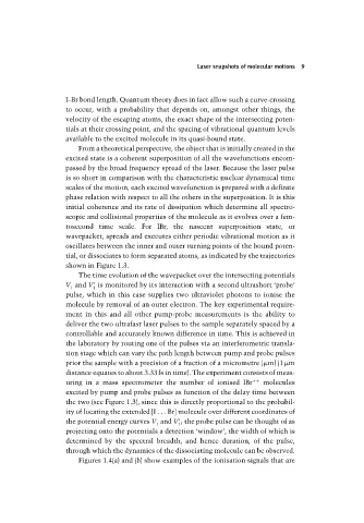
shown in Figure 1.3.
The time evolution of the wavepacket over the intersecting potentials
V and V is monitored by its interaction with a second ultrashort ‘probe’
1 1
pulse, which in this case supplies two ultraviolet photons to ionise the
molecule by removal of an outer electron. The key experimental require-
ment in this and all other pump-probe measurements is the ability to
deliver the two ultrafast laser pulses to the sample separately spaced by a
controllable and accurately known difference in time. This is achieved in
the laboratory by routing one of the pulses via an interferometric transla-
tion stage which can vary the path length between pump and probe pulses
prior the sample with a precision of a fraction of a micrometre ( m) (1 m
distance equates to about 3.33fs in time). The experiment consists of meas-
uring in a mass spectrometer the number of ionised IBr* molecules
excited by pump and probe pulses as function of the delay time between
the two (see Figure 1.3), since this is directly proportional to the probabil-
ity of locating the extended [I . . . Br] molecule over different coordinates of
the potential energy curves V and V ; the probe pulse can be thought of as
1 1
projecting onto the potentials a detection ‘window’, the width of which is
determined by the spectral breadth, and hence duration, of the pulse,
through which the dynamics of the dissociating molecule can be observed.
Figures 1.4(a) and (b) show examples of the ionisation signals that are