
Page 146 - Advances in Biomechanics and Tissue Regeneration
P. 146
142 8. TOWARDS THE REAL-TIME MODELING OF THE HEART
8.2.1 Passive Stress
The heart muscle tissue is adequately modeled by an exponential function coupled with an incompressibility term,
as suggested by Usyk and McCulloch [49]. This nonlinear orthotropic hyperelastic strain energy function has been
reformulated in terms of the invariants of the Green strain tensor, E, by Legner et al. [50] and is used in this research:
A Q
W ¼ ðe 1Þ + A comp ½detJlnðdetJÞ detJ +1, (8.1)
2
where
2 2 2
(8.2)
Q ¼ a 1 ðtrðM 1 EÞÞ + a 2 ðtrðM 2 EÞÞ + a 3 ðtrðM 3 EÞÞ
2
2
2
+ a 4 ðtrðM 1 EÞ Þ + a 5 ðtrðM 5 EÞ Þ + a 6 ðtrðM 3 EÞ Þ:
(8.3)
E ¼ E 11 V 1
V 1 + E 22 V 2
V 2 + E 33 V 3
V 3 + E 12 ðV 1
V 2 + V 2
V 1 Þ
+ E 13 ðV 1
V 3 + V 3
V 1 Þ + E 23 ðV 2
V 3 + V 3
V 2 Þ:
M 1 ¼ V 1
V 1 , M 2 ¼ V 2
V 2 , M 3 ¼ V 3
V 3 : (8.4)
J ¼ detF is the Jacobian with F denoting the deformation gradient tensor and A comp is a penalty parameter to control the
degree of incompressibility of cardiac tissue. The material constant A is a stress-scaling factor and a i (i ¼ 1, …, 6) are the
anisotropy coefficients associated with the local components of E corresponding to the preferred material directions,
namely fiber axis, V 1 , sheet axis, V 2 , and sheet normal axis, V 3 .
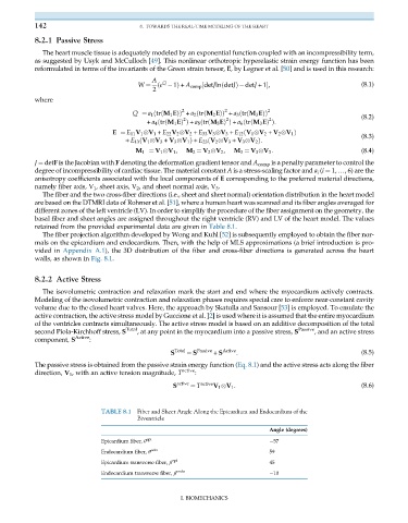
The fiber and the two cross-fiber directions (i.e., sheet and sheet normal) orientation distribution in the heart model
are based on the DTMRI data of Rohmer et al. [51], where a human heart was scanned and its fiber angles averaged for
different zones of the left ventricle (LV). In order to simplify the procedure of the fiber assignment on the geometry, the
basal fiber and sheet angles are assigned throughout the right ventricle (RV) and LV of the heart model. The values
retained from the provided experimental data are given in Table 8.1.
The fiber projection algorithm developed by Wong and Kuhl [52] is subsequently employed to obtain the fiber nor-
mals on the epicardium and endocardium. Then, with the help of MLS approximations (a brief introduction is pro-
vided in Appendix A.1), the 3D distribution of the fiber and cross-fiber directions is generated across the heart
walls, as shown in Fig. 8.1.
8.2.2 Active Stress
The isovolumetric contraction and relaxation mark the start and end where the myocardium actively contracts.
Modeling of the isovolumetric contraction and relaxation phases requires special care to enforce near-constant cavity
volume due to the closed heart valves. Here, the approach by Skatulla and Sansour [53] is employed. To emulate the
active contraction, the active stress model by Guccione et al. [2] is used where it is assumed that the entire myocardium
of the ventricles contracts simultaneously. The active stress model is based on an additive decomposition of the total
second Piola-Kirchhoff stress, S Total , at any point in the myocardium into a passive stress, S Passive , and an active stress
component, S Active :
S Total ¼ S Passive + S Active : (8.5)
The passive stress is obtained from the passive strain energy function (Eq. 8.1) and the active stress acts along the fiber
direction, V 1 , with an active tension magnitude, T active :
S active ¼ T active V 1
V 1 : (8.6)
TABLE 8.1 Fiber and Sheet Angle Along the Epicardium and Endocardium of the
Biventricle
Angle (degrees)
Epicardium fiber, θ epi 57
Endocardium fiber, θ edo 59
Epicardium transverse fiber, β epi 45
Endocardium transverse fiber, β endo 10
I. BIOMECHANICS