
Page 190 - Advances in Biomechanics and Tissue Regeneration
P. 190
186 9. COMPUTATIONAL MUSCULOSKELETAL BIOMECHANICS OF THE KNEE JOINT
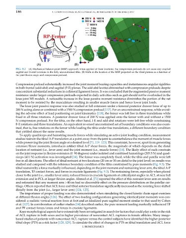
FIG. 9.2 (A) Mechanical balance point (MBP) approach; when applied at these locations, the compression preloads do not cause any coupled
sagittal and frontal rotations in the fully unconstrained tibia. (B) Shifts in the location of the MBP projected on the tibial plateau as a function of
the joint flexion angle and compression preload.
Compression preload substantially increased the joint moment bearing capacities and instantaneous angular rigidities
in both frontal (add-abd) and sagittal (F-E) planes. The add-abd laxities diminished with compression preloads despite
concomitant substantial reductions in collateral ligament forces. It was concluded that the augmented passive moment
resistance under larger compression preloads expected in daily activities such as gait should not be overlooked in the
knee joint MS models. A noticeable increase in the knee passive moment resistance diminishes the portion of the net
moment to be resisted by the musculature resulting in smaller muscle forces and hence lower joint loads.
The knee joint passive response was also studied at full extension under a femoral posterior drawer force of up to
200 N acting alone or combined with a 1500 N compression preload [117]. For an unconstrained response, while avoid-
ing the adverse effect of load positioning on joint kinematics [118], the femur was left free in three translations while
fixed in all three rotations. A posterior drawer force of 200 N was applied onto the femur with and without a 1500
N compression preload. For the tibia, on the other hand, I-E and add-abd rotations were left free while constraining
F-E rotations and three translations. An equivalent reversed unconstrained set of boundary conditions was also exam-
ined, that is, free rotations on the femur while loading the tibia under free translations, a different boundary condition
that yielded almost the same results.
To apply quadriceps and hamstring muscle forces while simulating an active joint loading condition, measurement
studies restrain the tibial A-P translation at a point away from the joint to counterbalance the moment of muscle forces
while preserving the joint flexion angle at a desired level [71, 119–123]. This constraint, however, apart from generating
extensor/flexor moments, introduces artifact tibial A-P shear forces, the magnitude of which depends on the distal
location of restraint (i.e., lever arm) and the joint moment (i.e., muscle forces) [14]. The likely effect of such constrain
on the joint response in flexion-extension (0–90 degrees) under isolated and combined hamstrings (205.5 N) and quad-
riceps (411 N) activation was investigated [14]. The femur was completely fixed, while the tibia and patella were left
free in all directions. The effect of tibial restraint at two locations (20 cm or 30 cm distal to the joint level) on results was
studied and compared with the reference boundary condition of the tibia constrained by pure moments (Fig. 9.3). The
tibial restraint by a force markedly influenced, depending on the joint moment and restraining lever arm, the tibial A-P
translation, TF contact forces, and forces in cruciate ligaments (Fig. 9.3). The restraining forces, especially when placed
closer to the joint (i.e., smaller lever arm), reduced forces in cruciate ligaments at critical joint angles: in ACL at near full
extension and in PCL at larger flexion angles. Ahmed et al. [71] reported the effect of this restraint lever arm on results
and estimated that any moment arm >40cm had no discernible effect on the pressure distribution on the patellar car-
tilage. Others reported that ACL force and tibial anterior translation significantly increased as the resisting force shifted
distally from the joint (i.e., larger lever arm) [124, 125].
The importance of proper loading was also demonstrated when simulating the closed kinetic chain squat exercises
at different flexion angles [126]. The effect of two loading configurations generating identical joint moments was con-
sidered: a realistic vertical reaction force at foot and an idealized pure sagittal moment similar to that used by Cohen
et al. [127]. In corroboration of earlier studies [14] described earlier, the pure moment loading markedly influenced TF
and PF contact forces/areas and forces in cruciate ligaments.
Knee morphological aspects have extensively been investigated in search for factors that could play a role in the risk
of ACL rupture in both sexes and in higher prevalence of noncontact ACL ruptures in female athletes. Many image-
based studies of patients with noncontact ACL rupture versus the control subjects have identified the higher posterior
tibial slope (PTS) as a risk factor [128, 129]. To simulate the effect of changes in PTS on tibial translation and ACL force
I. BIOMECHANICS