
Page 188 - Advances in Biomechanics and Tissue Regeneration
P. 188
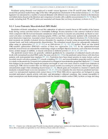
184                     9. COMPUTATIONAL MUSCULOSKELETAL BIOMECHANICS OF THE KNEE JOINT

              Nonlinear spring elements were employed to model various ligaments of the TF and PF joints. MCL wrapped
           around the proximal medial bony edge of the tibia with peripheral attachments to the medial meniscus [11, 81]. Each
           ligament was simulated by a number of uniaxial elements with different cross-sectional areas (based on the literature)
           and initial strains (based on the literature and comparison of results with available measurements) [82, 94–96]. Knee FE
           model, including both TF and PF joints and associated soft tissues (but not bony structures), is shown in Fig. 9.1.


           9.2.3 Lower Extremity Musculoskeletal (MS) Model
              Resolution of kinetic redundancy toward the estimation of unknown muscle forces in MS models of the human
           body during various activities remains a formidable challenge. Inverse dynamics is the common method of choice
           when compared with the forward dynamic simulations where activity in muscles are prescribed, say based on mea-
           sured activation via limited surface electromyography (EMG), and then continuously updated with constraints on
           some kinematics trajectories, measured contact forces, joint moments, and/or objective functions [30]. In the former,
           joint moments are initially evaluated (inverse dynamics) by equations of motion using measured joint kinematics,
           external loads, and body anthropometric characteristics. The redundant muscle forces are subsequently estimated,
           either using an optimization [21, 25, 97–101] or an EMG-driven [17, 18, 102–105] method. There are also hybrid
           EMG-assisted optimization (EMGAO) versions of these two approaches [106, 107]. In the optimization-based
           methods, muscle forces are estimated by minimizing a single or multiple objective functions, such as the sum of muscle
           forces, system margin of stability, or muscle activations to different powers [15, 20, 21, 25, 108–110].
              The predicted muscle forces are generally validated qualitatively by comparison of estimated muscle activation
           levels with normalized recorded EMG under the same activity [20, 110] or the predicted contact forces with data from
           patients with instrumented knee implants [97, 101]. Predictions have been found sensitive to many factors, such as
           recorded muscle activation patterns [97], muscle weighting [99, 101], and musculotendon properties and lever arms.
           In a study on the sensitivity of muscle force estimations to changes in musculotendon properties, Redl et al. [111] found
           that changes in the muscle fiber length and tendon rest length of vasti were most critical to model force estimates in
           normal gait. EMG-driven models often use Hill-type muscle models (accounting for activation, fiber length/velocity,
           and pennation angle) when estimating muscle forces [17, 31]. Muscle gains are evaluated in a manner to match joint
           moments evaluated based on inverse dynamics [17, 18, 27, 102] or to minimize the error between predicted and mea-
           sured (via instrumented implants) joint contact forces [103]. While EMG-driven approaches are biological in using
           recorded individual’s muscle activity with inter- and intrasubject volitional variations, they remain susceptible to
           major assumptions and shortcomings associated with the limited available surface EMG, location on large and deep


           FIG. 9.1  Posterior view of the knee joint FE model showing articular cartilage layers, menisci, and major ligaments. Rigid bony structures (i.e.,
           femur, tibia, and patella) are represented by their reference primary nodes and not shown.

                                                       I. BIOMECHANICS