
Page 236 - Advances in Biomechanics and Tissue Regeneration
P. 236
232 11. ANALYSIS OF THE BIOMECHANICAL BEHAVIOR OF INTRAMEDULLARY NAILING
U. U3 U. U3
0.19 0.22
0.14 0.17
0.10 0.11
0.05 0.06
0.00 0.01
–0.05 –0.04
–0.09 –0.10
–0.14 –0.15
–0.19 –0.20
–0.24 –0.26
–0.28 –0.31
–0.33 –0.36
–0.38 –0.41
(A) (B)
U. U3
U. U3 0.19
0.23 0.14
0.17 0.10
0.12 0.05
0.06 0.00
0.01 –0.05
–0.04 –0.09
–0.10 –0.14
–0.15 –0.19
–0.21 –0.24
–0.26 –0.28
–0.31 –0.33
–0.37 –0.38
–0.42
(C) (D)
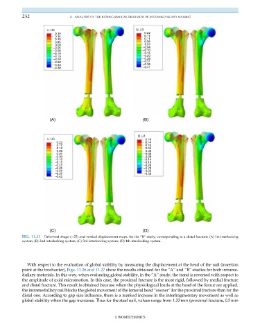
FIG. 11.23 Deformed shape ( 25) and vertical displacement maps, for the “B” study, corresponding to a distal fracture: (A) 1st interlocking
system; (B) 2nd interlocking system; (C) 3rd interlocking system; (D) 4th interlocking system.
With respect to the evaluation of global stability by measuring the displacement at the head of the nail (insertion
point at the trochanter), Figs. 11.26 and 11.27 show the results obtained for the “A” and “B” studies for both intrame-
dullary materials. In this way, when evaluating global stability, in the “A” study, the trend is reversed with respect to
the amplitude of axial micromotion. In this case, the proximal fracture is the most rigid, followed by medial fracture
and distal fracture. This result is obtained because when the physiological loads at the head of the femur are applied,
the intramedullary nail blocks the global movement of the femoral head “sooner” for the proximal fracture than for the
distal one. According to gap size influence, there is a marked increase in the interfragmentary movement as well as
global stability when the gap increases. Thus for the steel nail, values range from 1.33mm (proximal fracture, 0.5mm
I. BIOMECHANICS