
Page 233 - Advances in Biomechanics and Tissue Regeneration
P. 233
11.3 TYPES OF FRACTURES AND OSTEOSYNTHESIS 229
to an analysis of the biomechanical behavior of different systems of osteosynthesis (with different locking systems) for
different types of fracture with the same location in the femur (distal).
All the considered fractures were modeled as transverse by means of an irregular surface developed to represent a
closer geometry to the actual fracture. The effect of the gap size was unclear in the literature. So, the majority of the
reviewed in vivo studies referred to a gap size ranging from 0.6 to 6mm [40, 50], whereas in FE simulation articles it
ranged from 0.7 to 10mm [47, 51].
Thus for the “A” study, three different fracture gaps were studied: 0.5mm (considered as a noncomminuted frac-
ture), 3mm (the most referenced value found in the literature, representing a mid-value), and 20mm as an example of a
comminuted fracture. In addition to this, three localizations of the fracture were studied: proximal, medial, and distal
for each gap size. Only one combination of screws was studied: one oblique placed proximally and two transversely at
the distal part. On the other hand, the purpose of the “B” study was to investigate the optimal screw combination and
gap size for a single distal fracture location, considering the same three gap sizes: 0.5, 3, and 20mm, respectively. Thus
four combinations of locking screws were considered: one oblique proximal screw combined with four configurations
of the three distal ones, two lateral-medial and one anteroposterior. Table 11.3 summarizes the list of FE models sim-
ulated for the “A” and “B” studies (9 and 12 FE models, respectively). These models will be duplicated, since each one
of these cases is carried out considering the two studied materials (stainless steel and titanium) of the nail.
Finally, to validate the conclusions obtained from simulations, a clinical follow-up was carried out for both studies,
approved by the Ethics Committee of the Institute of Health Sciences of Aragón (protocol number CI PI15/0214). Thus
for the “A” study, a sample of 55 patients, 24 males and 31 females, with a mean age of 52.5years was obtained, all of
them treated with femoral nail Stryker S2. Localizations of fractures were 32 in the right femur and 33 in the left femur.
On the other hand, for the “B” study, a sample of 15 patients, 6 males and 9 females, with a mean age of 53.2years was
obtained, all of them treated with the same nail as in the “A” study. Localizations of fractures were 10 in the right femur
and 5 in the left femur. The grade of comminution was measured in both cases according to the scale of Winquist and
Hansen [5]. The distribution of cases corresponding to fracture localization and fracture grade are included, for the “A”
and “B” studies, in Table 11.4.
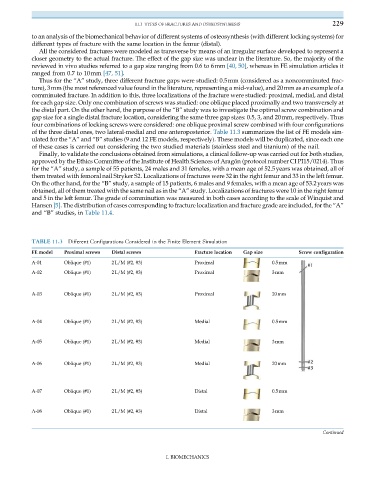
TABLE 11.3 Different Configurations Considered in the Finite Element Simulation
FE model Proximal screws Distal screws Fracture location Gap size Screw configuration
A-01 Oblique (#1) 2L/M (#2, #3) Proximal 0.5mm
A-02 Oblique (#1) 2L/M (#2, #3) Proximal 3mm
A-03 Oblique (#1) 2L/M (#2, #3) Proximal 20mm
A-04 Oblique (#1) 2L/M (#2, #3) Medial 0.5mm
A-05 Oblique (#1) 2L/M (#2, #3) Medial 3mm
A-06 Oblique (#1) 2L/M (#2, #3) Medial 20mm
A-07 Oblique (#1) 2L/M (#2, #3) Distal 0.5mm
A-08 Oblique (#1) 2L/M (#2, #3) Distal 3mm
Continued
I. BIOMECHANICS