
Page 238 - Advances in Biomechanics and Tissue Regeneration
P. 238
234 11. ANALYSIS OF THE BIOMECHANICAL BEHAVIOR OF INTRAMEDULLARY NAILING
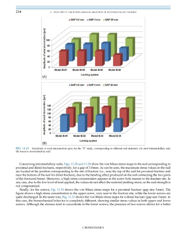
FIG. 11.25 Amplitude of axial micromotion (μm), for the “B” study, corresponding to different nail materials: (A) steel intramedullary nail;
(B) titanium intramedullary nail.
Concerning intramedullary nails, Figs. 11.28 and 11.29 show the von Mises stress maps in the nail corresponding to
proximal and distal fractures, respectively, for a gap of 3.0mm. As can be seen, the maximum stress values in the nail
are located at the position corresponding to the site of fracture (i.e., near the top of the nail for proximal fracture and
near the bottom of the nail for distal fracture), due to the bending effect produced on the nail connecting the two parts
of the fractured femur. Moreover, a high stress concentration appears in the screw hole nearest to the fracture site. In
any case, due to the low level of load applied, the values do not affect the material yielding stress, so the nail strength is
not compromised.
Finally, for the screws, Fig. 11.30 shows the von Mises stress maps for a proximal fracture (gap size 3mm). The
figure shows a high stress concentration in the upper screw, very near to the fracture site, while the lower screws are
quite discharged. In the same way, Fig. 11.31 shows the von Mises stress maps for a distal fracture (gap size 3mm). In
this case, the biomechanical behavior is completely different, showing similar stress values in both upper and lower
screws. Although the stresses tend to concentrate in the lower screws, the presence of two screws allows for a better
I. BIOMECHANICS