
Page 291 - Advances in Biomechanics and Tissue Regeneration
P. 291
15.1 INTRODUCTION 289
migration, at least occasionally [21]. In the presence of other cells, the cell-cell attraction may affect cell-ECM adhesion
and may facilitate cell-cell contacts. Hence, the dynamics of collective cell migration result in complex changes in multi-
cellular tissue structures. Disparate single cell migration, collective cell migration serves to keep the tissue intact during
remodeling. The cell-cell interactions and cell motility coordination during multicell migration can be studied from
two perspectives. First, how do the cells affect each other? To what radius does a cell transmit force and communicate
for transmitting information? Second, how can cell-cell interactions affect their individual and collective behavior?
Understanding these perspectives can help to answer many questions such as Does collective cell migration speed
up or delay cell movement? How do cell slugs affect each other? Some of these questions may be answered via exper-
imental works, but to profoundly answer these questions and much more, numerical studies are inevitable.
Cell migration on two-dimensional (2D) surfaces takes place during the reepithelialization of wounds, the scanning
of leukocytes along the inner blood vessel wall, or inner epithelial surfaces [21]. Although 2D studies have enhanced
our insights into many contexts such as the basic mechanisms by which cells migrate, interact with the ECM, and
change their speed or direction, they may sometimes impose an artificial apical-based cell polarity that may not exist
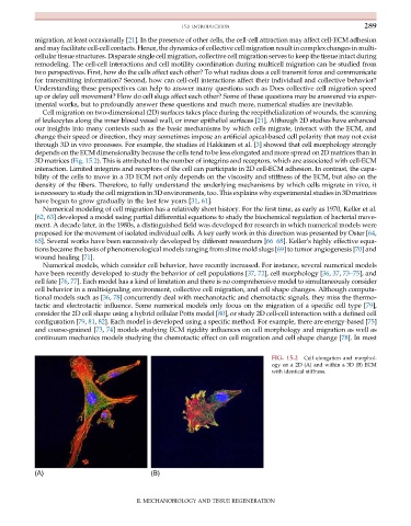
through 3D in vivo processes. For example, the studies of Hakkinen et al. [3] showed that cell morphology strongly
depends on the ECM dimensionality because the cells tend to be less elongated and more spread on 2D matrices than in
3D matrices (Fig. 15.2). This is attributed to the number of integrins and receptors, which are associated with cell-ECM
interaction. Limited integrins and receptors of the cell can participate in 2D cell-ECM adhesion. In contrast, the capa-
bility of the cells to move in a 3D ECM not only depends on the viscosity and stiffness of the ECM, but also on the
density of the fibers. Therefore, to fully understand the underlying mechanisms by which cells migrate in vivo, it
is necessary to study the cell migration in 3D environments, too. This explains why experimental studies in 3D matrices
have begun to grow gradually in the last few years [31, 61].
Numerical modeling of cell migration has a relatively short history. For the first time, as early as 1970, Keller et al.
[62, 63] developed a model using partial differential equations to study the biochemical regulation of bacterial move-
ment. A decade later, in the 1980s, a distinguished field was developed for research in which numerical models were
proposed for the movement of isolated individual cells. A key early work in this direction was presented by Oster [64,
65]. Several works have been successively developed by different researchers [66–68]. Keller’s highly effective equa-
tions became the basis of phenomenological models ranging from slime mold slugs [69] to tumor angiogenesis [70] and
wound healing [71].
Numerical models, which consider cell behavior, have recently increased. For instance, several numerical models
have been recently developed to study the behavior of cell populations [37, 72], cell morphology [36, 37, 73–75], and
cell fate [76, 77]. Each model has a kind of limitation and there is no comprehensive model to simultaneously consider
cell behavior in a multisignaling environment, collective cell migration, and cell shape changes. Although computa-
tional models such as [36, 78] concurrently deal with mechanotactic and chemotactic signals, they miss the thermo-
tactic and electrotactic influence. Some numerical models only focus on the migration of a specific cell type [79],
consider the 2D cell shape using a hybrid cellular Potts model [80], or study 2D cell-cell interaction with a defined cell
configuration [79, 81, 82]. Each model is developed using a specific method. For example, there are energy-based [75]
and coarse-grained [73, 74] models studying ECM rigidity influences on cell morphology and migration as well as
continuum mechanics models studying the chemotactic effect on cell migration and cell shape change [78]. In most
FIG. 15.2 Cell elongation and morphol-
ogy on a 2D (A) and within a 3D (B) ECM
with identical stiffness.
(A) (B)
II. MECHANOBIOLOGY AND TISSUE REGENERATION