
Page 290 - Advances in Biomechanics and Tissue Regeneration
P. 290
288 15. COMPUTATIONAL SIMULATION OF CELL BEHAVIOR FOR TISSUE REGENERATION
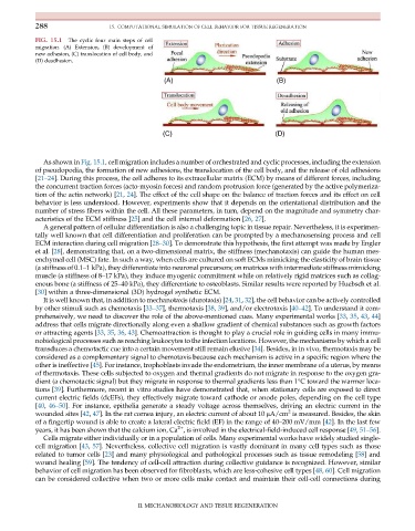
FIG. 15.1 The cyclic four main steps of cell
migration. (A) Extension, (B) development of
new adhesion, (C) translocation of cell body, and
(D) deadhesion.
(A) (B)
(C) (D)
As shown in Fig. 15.1, cell migration includes a number of orchestrated and cyclic processes, including the extension
of pseudopodia, the formation of new adhesions, the translocation of the cell body, and the release of old adhesions
[21–24]. During this process, the cell adheres to its extracellular matrix (ECM) by means of different forces, including
the concurrent traction forces (acto-myosin forces) and random protrusion force (generated by the active polymeriza-
tion of the actin network) [21, 24]. The effect of the cell shape on the balance of traction forces and its effect on cell
behavior is less understood. However, experiments show that it depends on the orientational distribution and the
number of stress fibers within the cell. All these parameters, in turn, depend on the magnitude and symmetry char-
acteristics of the ECM stiffness [25] and the cell internal deformation [26, 27].
A general pattern of cellular differentiation is also a challenging topic in tissue repair. Nevertheless, it is experimen-
tally well known that cell differentiation and proliferation can be prompted by a mechanosensing process and cell
ECM interaction during cell migration [28–30]. To demonstrate this hypothesis, the first attempt was made by Engler
et al. [28], demonstrating that, on a two-dimensional matrix, the stiffness (mechanotaxis) can guide the human mes-
enchymal cell (MSC) fate. In such a way, when cells are cultured on soft ECMs mimicking the elasticity of brain tissue
(a stiffness of 0.1–1 kPa), they differentiate into neuronal precursors; on matrices with intermediate stiffness mimicking
muscle (a stiffness of 8–17 kPa), they induce myogenic commitment while on relatively rigid matrices such as collag-
enous bone (a stiffness of 25–40 kPa), they differentiate to osteoblasts. Similar results were reported by Huebsch et al.
[30] within a three-dimensional (3D) hydrogel synthetic ECM.
It is well known that, in addition to mechanotaxis (durotaxis) [24, 31, 32], the cell behavior can be actively controlled
by other stimuli such as chemotaxis [33–37], thermotaxis [38, 39], and/or electrotaxis [40–42]. To understand it com-
prehensively, we need to discover the role of the above-mentioned cues. Many experimental works [33, 35, 43, 44]
address that cells migrate directionally along even a shallow gradient of chemical substances such as growth factors
or attracting agents [33, 35, 36, 43]. Chemoattraction is thought to play a crucial role in guiding cells in many immu-
nobiological processes such as reaching leukocytes to the infection locations. However, the mechanisms by which a cell
transduces a chemotactic cue into a certain movement still remain elusive [34]. Besides, in in vivo, thermotaxis may be
considered as a complementary signal to chemotaxis because each mechanism is active in a specific region where the
other is ineffective [45]. For instance, trophoblasts invade the endometrium, the inner membrane of a uterus, by means
of thermotaxis. These cells subjected to oxygen and thermal gradients do not migrate in response to the oxygen gra-
dient (a chemotactic signal) but they migrate in response to thermal gradients less than 1°C toward the warmer loca-
tions [39]. Furthermore, recent in vitro studies have demonstrated that, when stationary cells are exposed to direct
current electric fields (dcEFs), they effectively migrate toward cathode or anode poles, depending on the cell type
[40, 46–50]. For instance, epithelia generate a steady voltage across themselves, driving an electric current in the
2
wounded sites [42, 47]. In the rat cornea injury, an electric current of about 10 μA/cm is measured. Besides, the skin
of a fingertip wound is able to create a lateral electric field (EF) in the range of 40–200 mV/mm [42]. In the last few
2+
years, it has been shown that the calcium ion, Ca , is involved in the electrical-field-induced cell response [49, 51–56].
Cells migrate either individually or in a population of cells. Many experimental works have widely studied single-
cell migration [43, 57]. Nevertheless, collective cell migration is vastly dominant in many cell types such as those
related to tumor cells [23] and many physiological and pathological processes such as tissue remodeling [58] and
wound healing [59]. The tendency of cell-cell attraction during collective guidance is recognized. However, similar
behavior of cell migration has been observed for fibroblasts, which are less-cohesive cell types [48, 60]. Cell migration
can be considered collective when two or more cells make contact and maintain their cell-cell connections during
II. MECHANOBIOLOGY AND TISSUE REGENERATION