
Page 293 - Advances in Biomechanics and Tissue Regeneration
P. 293
15.2 METHODOLOGY 291
these effective forces on the cell body should be in equilibrium with the opposing drag force. To predict the cell
response to multiple signals received from the cell environment, we developed a 3D computational model using a
discrete finite element methodology considering the cell as a group of finite elements. It is considered that ECM
and the cell constitute the working domain. The working domain is divided into a number of subdomains (depending
on the cell number) and each subdomain is considered to represent a cell. The cell behavior is modeled using two dif-
ferent strategies:
1. Constant spherical cell morphology to investigate cell migration, differentiation, and proliferation in the presence of
different stimuli.
2. Variable cell morphology to study the cell morphology in the presence of different stimuli.
This allows us to predict the cell behavior and response when it is surrounded by different microenvironmental
physical characteristics.
15.2.1 Mechanotaxis
The presented model can be employed to simulate adherent cells that are cultured on 2D or within 3D ECM. Cells
have a special internal structure that is prepared to detect the stiffness of the matrix in which they reside. For instance,
fibroblasts preferentially move toward stiffer ECMs [26, 96]. This phenomenon is known as mechanotaxis by which a
cell moves directionally toward stiffer regions within its ECM [97]. In the mechanosensing step, the cell senses its ECM
by exerting a sensing force to diagnose its microenvironment and to obtain information about its ECM rigidity. When
the cell determines its surrounding mechanical conditions, it starts to pull itself toward the stiffer and/or more
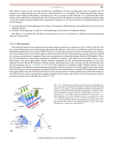
fixed region. The active apparatuses cellular elements regulating the cell mechanosensing process are the actin
filaments and the Myosin II machinery and the passive mechanical ones of the cell body are the microtubules and
the cell membrane (see Fig. 15.3A)[26, 32, 72, 97, 98]. The cytoplasmic cytoskeletal (CSK) is linked with the external
ECM through focal adhesions and transmembrane integrins that are assumed to be totally rigid in the present model.
This scheme agrees with the tensegrity hypothesis [96] because the deformation of external ECM is balanced by tensile
forces generated in the actin CSK. External forces are also considered to be another possible cause of the deformation of
the ECM and cell. During cell migration, certain mechanical forces such as the traction force, the drag force, and the
protrusion random force will affect the cell [24, 97, 99].
FIG. 15.3 Mechanosensing model of an adherent cell. (A) Schematic dia-
gram of the relevant mechanical constituents of a cell [26]. (B) Cell mechanical
model. K act , K pas , and K subs denote the stiffness modulus of actin filaments, the
passive components of the cell, and the ECM, respectively. f ext stands for exter-
act
nal forces applied to the cell or the ECM. (C) Dependence of the active, σ ,
and passive, σ pas , contractile stresses on the deformation of the cell, E. E act is
the deformation of the active contractile elements. σ max stands for the maxi-
mum contractile stress exerted by the actin-myosin machinery, and E min and
E max are the corresponding shortening and lengthening strains of the contrac-
tile elements with respect to the unloaded length at which active stress
becomes zero. σ s is the stress of the ECM [26].
(A)
(B) (C)
II. MECHANOBIOLOGY AND TISSUE REGENERATION