Page 297 - Advances in Biomechanics and Tissue Regeneration
P. 297
15.2 METHODOLOGY 295
0:09
l max l med
(15.13)
l min
f shape ¼ 2
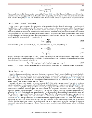
This is nearly identical to the expression proposed by Dressel [114], except that he used a 0.1 exponent. Other shape
factors have also been suggested to characterize the shape irregularity, but the impact of the max-med-min area factor
tends to be the strongest [112, 115]. It is notable that this shape factor in the case of a spherical cell shape delivers one.
15.2.2 Chemotaxis and Thermotaxis
In the presence of chemotaxis or thermotaxis, the cell polarization direction depends not only on the mechanotactic
signal, but also on those additional stimuli. We assume that the presence of these cues changes neither the properties of
a typical cell nor its surrounding ECM. Because the exerted traction forces by the cell depend on the cell type and the
mechanical properties of the ECM, the presence of these cues does not affect the magnitude of the net traction force but
changes its direction. The realignment of the net traction force in the presence of chemical and thermal cues changes
based on the direction of chemotaxis and thermotaxis gradients. Therefore, it is assumed that a part of the net traction
force is guided by mechanotaxis by a unit vector of
F trac
net
k F
e mech ¼ trac (15.14)
net k
while the rest is guided by chemotaxis, e ch , and/or thermotaxis, e th , cues, respectively, as
(15.15)
r½C
e ch ¼
kr½Ck
rT
(15.16)
e th ¼
krT k
where r is the gradient operator and [C] and T are the chemoattractant concentration and the temperature, respec-
tively. Consequently, based on each cue, the effective direction on the cell and the effective force due to mechanotaxis,
chemotaxis, and thermotaxis is calculated as
e
F eff ¼ F trac ðμ mech mech + μ e ch + μ e th Þ; μ + μ + μ ¼ 1 (15.17)
net ch th mech ch th
where μ mech , μ ch , and μ th are the efficient factors of mechanotaxis, chemotaxis, and thermotaxis cues, respectively.
15.2.3 Electrotaxis
Based on the experimental observations, the electrotactic response of the cell is controlled by an intracellular influx
of Ca 2+ [47, 48]. However, it is still a controversial question. For instance, Ca 2+ dependence of electrotaxis has been
observed in neural crest cells, embryo mouse fibroblasts, and fish and human keratocytes [40, 47, 49, 60, 116]. By con-
trast, Ca 2+ independent electrotaxis has been reported in mouse fibroblasts [51]. Therefore, the precise mechanism
behind the Ca 2+ role in electrotaxis is not well known.
A simple cell in rest has a negative membrane potential [47]. If this cell is exposed to a dcEF, the side of its plasma
membrane that faces the cathode depolarizes while the other side that faces the anode hyperpolarizes [40, 47, 60]. In the
case of a cell with negligible voltage-gated conductance, the hyperpolarized membrane side attracts Ca 2+ by passive
electrochemical diffusion. This side of the cell, then, contracts and propels the cell toward the cathode. This process
continues until the voltage-gated Ca 2+ channels (VGCCs) near the cathodal side open (depolarized) to allow Ca 2+
influx (see Fig. 15.6). Consequently, the intracellular Ca 2+ level rises on both the anodal and cathodal sides of the cell.
The resultant of the generated force, in this case, would depend on the balance between the opposing magnetic con-
tractile forces [47]. That is why some cells reorient toward the cathode, such as embryo fibroblasts [49], human ker-
atinocytes [48, 117], fish epidermal cells [116], and human retinal pigment epithelial cells [118], while some others do so
toward the anode, such as human granulocytes [119] and metastatic human breast cancer cells [120]. To define the
effective electrical force in the presence of an electrotactic cue, let us assume a cell located in a uniform EF. As a con-
sequence, it is ionized and has acquired a charge. The experienced force by this individual charged cell can be obtained
by [121]
II. MECHANOBIOLOGY AND TISSUE REGENERATION

